 Movie
Movie Controller


+ Open data

- Basic information
| Entry | Database: PDB / ID: 6pj6 | ||||||||||||
|---|---|---|---|---|---|---|---|---|---|---|---|---|---|
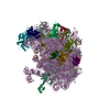
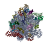
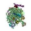
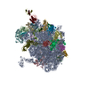
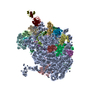
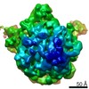
| Title | High resolution cryo-EM structure of E.coli 50S | ||||||||||||
 Components Components |
| ||||||||||||
 Keywords Keywords | RIBOSOME | ||||||||||||
| Function / homology |  Function and homology information Function and homology informationribosomal large subunit assembly / large ribosomal subunit / transferase activity / 5S rRNA binding / cytosolic large ribosomal subunit / cytoplasmic translation / tRNA binding / rRNA binding / ribosome / structural constituent of ribosome ...ribosomal large subunit assembly / large ribosomal subunit / transferase activity / 5S rRNA binding / cytosolic large ribosomal subunit / cytoplasmic translation / tRNA binding / rRNA binding / ribosome / structural constituent of ribosome / translation / ribonucleoprotein complex / cytoplasm / cytosol Similarity search - Function | ||||||||||||
| Biological species |  | ||||||||||||
| Method | ELECTRON MICROSCOPY / single particle reconstruction / cryo EM / Resolution: 2.2 Å | ||||||||||||
 Authors Authors | Stojkovic, V. / Myasnikov, A. / Frost, A. / Fujimori, D.G. | ||||||||||||
| Funding support |  United States, 3items United States, 3items
| ||||||||||||
 Citation Citation | Journal: Nucleic Acids Res / Year: 2020 Title: Assessment of the nucleotide modifications in the high-resolution cryo-electron microscopy structure of the Escherichia coli 50S subunit. Authors: Vanja Stojković / Alexander G Myasnikov / Iris D Young / Adam Frost / James S Fraser / Danica Galonić Fujimori / Abstract: Post-transcriptional ribosomal RNA (rRNA) modifications are present in all organisms, but their exact functional roles and positions are yet to be fully characterized. Modified nucleotides have been ...Post-transcriptional ribosomal RNA (rRNA) modifications are present in all organisms, but their exact functional roles and positions are yet to be fully characterized. Modified nucleotides have been implicated in the stabilization of RNA structure and regulation of ribosome biogenesis and protein synthesis. In some instances, rRNA modifications can confer antibiotic resistance. High-resolution ribosome structures are thus necessary for precise determination of modified nucleotides' positions, a task that has previously been accomplished by X-ray crystallography. Here, we present a cryo-electron microscopy (cryo-EM) structure of the Escherichia coli 50S subunit at an average resolution of 2.2 Å as an additional approach for mapping modification sites. Our structure confirms known modifications present in 23S rRNA and additionally allows for localization of Mg2+ ions and their coordinated water molecules. Using our cryo-EM structure as a testbed, we developed a program for assessment of cryo-EM map quality. This program can be easily used on any RNA-containing cryo-EM structure, and an associated Coot plugin allows for visualization of validated modifications, making it highly accessible. | ||||||||||||
| History |
|
- Structure visualization
| Movie |
Movie viewer |
|---|---|
| Structure viewer | Molecule: MolmilJmol/JSmol |
- Downloads & links
-Download
| PDBx/mmCIF format | 6pj6.cif.gz | 3.2 MB | Display | PDBx/mmCIF format |
|---|---|---|---|---|
| PDB format | pdb6pj6.ent.gz | Display | PDB format | |
| PDBx/mmJSON format | 6pj6.json.gz | Tree view | PDBx/mmJSON format | |
| Others |  Other downloads Other downloads |
-Validation report
| Summary document | 6pj6_validation.pdf.gz | 1.3 MB | Display | wwPDB validaton report |
|---|---|---|---|---|
| Full document | 6pj6_full_validation.pdf.gz | 1.3 MB | Display | |
| Data in XML | 6pj6_validation.xml.gz | 132.3 KB | Display | |
| Data in CIF | 6pj6_validation.cif.gz | 250.3 KB | Display | |
| Arichive directory | https://data.pdbj.org/pub/pdb/validation_reports/pj/6pj6ftp://data.pdbj.org/pub/pdb/validation_reports/pj/6pj6 | HTTPS FTP |
-Related structure data
| Related structure data | 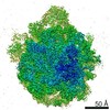 20353MC M: map data used to model this data C: citing same article ( |
|---|---|
| Similar structure data |
-Links
 PDBj
PDBj


- Assembly
| Deposited unit | 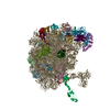
|
|---|---|
| 1 |
|
-Components
-RNA chain , 2 types, 2 molecules IJ
| #1: RNA chain | Mass: 941811.562 Da / Num. of mol.: 1 / Source method: isolated from a natural source / Source: (natural) |
|---|---|
| #2: RNA chain | Mass: 38177.762 Da / Num. of mol.: 1 / Source method: isolated from a natural source / Source: (natural) |
+50S ribosomal protein ... , 29 types, 29 molecules KLMNOPQRSTUVWXYZabcdefghijklm
-Non-polymers , 4 types, 4084 molecules 


| #32: Chemical | ChemComp-MG /  Mass: 24.305 Da / Num. of mol.: 185 / Source method: obtained synthetically / Formula: Mg Mass: 24.305 Da / Num. of mol.: 185 / Source method: obtained synthetically / Formula: Mg#33: Chemical | ChemComp-NA / |  Mass: 22.990 Da / Num. of mol.: 1 / Source method: obtained synthetically / Formula: Na Mass: 22.990 Da / Num. of mol.: 1 / Source method: obtained synthetically / Formula: Na#34: Chemical | ChemComp-ZN / |  Mass: 65.409 Da / Num. of mol.: 1 / Source method: obtained synthetically / Formula: Zn Mass: 65.409 Da / Num. of mol.: 1 / Source method: obtained synthetically / Formula: Zn#35: Water | ChemComp-HOH / | Mass: 18.015 Da / Num. of mol.: 3897 / Source method: isolated from a natural source / Formula: H2O |
|---|
-Experimental details
-Experiment
| Experiment | Method: ELECTRON MICROSCOPY |
|---|---|
| EM experiment | Aggregation state: PARTICLE / 3D reconstruction method: single particle reconstruction |
- Sample preparation
| Component | Name: Escherichia coli 50S subunit / Type: RIBOSOME / Entity ID: #1-#31 / Source: NATURAL |
|---|---|
| Source (natural) | Organism: |
| Buffer solution | pH: 7.5 / Details: Buffer A |
| Buffer component | Conc.: 10 mM / Name: TRIS / Formula: TRIS |
| Specimen | Conc.: 1 mg/ml / Embedding applied: NO / Shadowing applied: NO / Staining applied: NO / Vitrification applied: YES Details: 50S ribosomal subunit was purified from E. coli MRE600 using modified version of previously published protocol (Mehta et al. 2012) |
| Specimen support | Details: easyGlow settings / Grid material: COPPER / Grid mesh size: 300 divisions/in. / Grid type: Quantifoil R1.2/1.3 |
| Vitrification | Instrument: FEI VITROBOT MARK IV / Cryogen name: ETHANE / Humidity: 95 % / Chamber temperature: 10 K Details: Blot Force 5 Blot Time 10sec Hum 95% Temperature 10C Waiting time 1 min |
- Electron microscopy imaging
| Experimental equipment |  Model: Titan Krios / Image courtesy: FEI Company |
|---|---|
| Microscopy | Model: FEI TITAN KRIOS |
| Electron gun | Electron source:  FIELD EMISSION GUN / Accelerating voltage: 300 kV / Illumination mode: OTHER FIELD EMISSION GUN / Accelerating voltage: 300 kV / Illumination mode: OTHER |
| Electron lens | Mode: BRIGHT FIELD / Nominal magnification: 59000 X / Calibrated magnification: 60827 X / Nominal defocus max: 1200 nm / Nominal defocus min: 300 nm / Calibrated defocus min: 500 nm / Calibrated defocus max: 1500 nm / Cs: 2.7 mm / C2 aperture diameter: 70 µm / Alignment procedure: COMA FREE |
| Specimen holder | Cryogen: NITROGEN / Specimen holder model: FEI TITAN KRIOS AUTOGRID HOLDER / Temperature (max): 130 K / Temperature (min): 86 K / Residual tilt: 10 mradians |
| Image recording | Average exposure time: 8 sec. / Electron dose: 80 e/Å2 / Detector mode: SUPER-RESOLUTION / Film or detector model: GATAN K2 SUMMIT (4k x 4k) / Num. of grids imaged: 1 / Num. of real images: 1889 |
| Image scans | Sampling size: 5 µm / Width: 3838 / Height: 3710 / Movie frames/image: 80 / Used frames/image: 0-80 |
- Processing
| EM software |
| ||||||||||||||||||
|---|---|---|---|---|---|---|---|---|---|---|---|---|---|---|---|---|---|---|---|
| CTF correction | Type: NONE | ||||||||||||||||||
| Particle selection | Num. of particles selected: 193000 | ||||||||||||||||||
| Symmetry | Point symmetry: C1 (asymmetric) | ||||||||||||||||||
| 3D reconstruction | Resolution: 2.2 Å / Resolution method: FSC 0.143 CUT-OFF / Num. of particles: 141549 / Algorithm: FOURIER SPACE / Symmetry type: POINT |
