 Movie
Movie Controller


+ Open data

- Basic information
| Entry | Database: PDB / ID: 6pik | ||||||
|---|---|---|---|---|---|---|---|
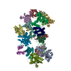
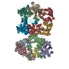
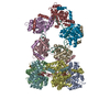
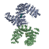
| Title | Tetrameric cryo-EM ArnA | ||||||
 Components Components |
| ||||||
 Keywords Keywords | ANTIBIOTIC / Polymyxin resistance / hexamer / tetramer | ||||||
| Function / homology |  Function and homology information Function and homology information: / UDP-4-deoxy-4-formamido-beta-L-arabinopyranose biosynthetic process / UDP-glucuronic acid dehydrogenase activity / UDP-4-amino-4-deoxy-L-arabinose formyltransferase activity / UDP-glucuronic acid dehydrogenase (UDP-4-keto-hexauronic acid decarboxylating) / UDP-4-amino-4-deoxy-L-arabinose formyltransferase / UDP-D-xylose biosynthetic process / UDP-glucuronate decarboxylase activity / lipopolysaccharide biosynthetic process / lipid A biosynthetic process ...: / UDP-4-deoxy-4-formamido-beta-L-arabinopyranose biosynthetic process / UDP-glucuronic acid dehydrogenase activity / UDP-4-amino-4-deoxy-L-arabinose formyltransferase activity / UDP-glucuronic acid dehydrogenase (UDP-4-keto-hexauronic acid decarboxylating) / UDP-4-amino-4-deoxy-L-arabinose formyltransferase / UDP-D-xylose biosynthetic process / UDP-glucuronate decarboxylase activity / lipopolysaccharide biosynthetic process / lipid A biosynthetic process / NAD+ binding / response to antibiotic / protein-containing complex / identical protein binding Similarity search - Function | ||||||
| Biological species |  | ||||||
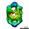
| Method | ELECTRON MICROSCOPY / single particle reconstruction / cryo EM / Resolution: 7.8 Å | ||||||
 Authors Authors | Yang, M. / Gehring, K. | ||||||
| Funding support |  Canada, 1items Canada, 1items
| ||||||
 Citation Citation | Journal: J Struct Biol / Year: 2019 Title: Cryo-electron microscopy structures of ArnA, a key enzyme for polymyxin resistance, revealed unexpected oligomerizations and domain movements. Authors: Meng Yang / Yu Seby Chen / Muneyoshi Ichikawa / Daniel Calles-Garcia / Kaustuv Basu / Rayan Fakih / Khanh Huy Bui / Kalle Gehring / Abstract: Gram-negative bacteria evade the attack of cationic antimicrobial peptides through modifying their lipid A structure in their outer membranes with 4-amino-4-deoxy-L-arabinose (Ara4N). ArnA is a ...Gram-negative bacteria evade the attack of cationic antimicrobial peptides through modifying their lipid A structure in their outer membranes with 4-amino-4-deoxy-L-arabinose (Ara4N). ArnA is a crucial enzyme in the lipid A modification pathway and its deletion abolishes the polymyxin resistance of gram-negative bacteria. Previous studies by X-ray crystallography have shown that full-length ArnA forms a three-bladed propeller-shaped hexamer. Here, the structures of ArnA determined by cryo-electron microscopy (cryo-EM) reveal that ArnA exists in two 3D architectures, hexamer and tetramer. This is the first observation of a tetrameric ArnA. The hexameric cryo-EM structure is similar to previous crystal structures but shows differences in domain movements and conformational changes. We propose that ArnA oligomeric states are in a dynamic equilibrium, where the hexamer state is energetically more favorable, and its domain movements are important for cooperating with downstream enzymes in the lipid A-Ara4N modification pathway. The results provide us with new possibilities to explore inhibitors targeting ArnA. | ||||||
| History |
|
- Structure visualization
| Movie |
Movie viewer |
|---|---|
| Structure viewer | Molecule: MolmilJmol/JSmol |
- Downloads & links
-Download
| PDBx/mmCIF format | 6pik.cif.gz | 473.7 KB | Display | PDBx/mmCIF format |
|---|---|---|---|---|
| PDB format | pdb6pik.ent.gz | 389.1 KB | Display | PDB format |
| PDBx/mmJSON format | 6pik.json.gz | Tree view | PDBx/mmJSON format | |
| Others |  Other downloads Other downloads |
-Validation report
| Summary document | 6pik_validation.pdf.gz | 1.1 MB | Display | wwPDB validaton report |
|---|---|---|---|---|
| Full document | 6pik_full_validation.pdf.gz | 1.1 MB | Display | |
| Data in XML | 6pik_validation.xml.gz | 77.5 KB | Display | |
| Data in CIF | 6pik_validation.cif.gz | 107.3 KB | Display | |
| Arichive directory | https://data.pdbj.org/pub/pdb/validation_reports/pi/6pikftp://data.pdbj.org/pub/pdb/validation_reports/pi/6pik | HTTPS FTP |
-Related structure data
| Related structure data |  20352MC  0456C 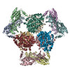 6pihC M: map data used to model this data C: citing same article ( |
|---|---|
| Similar structure data |

-Links
 PDBj
PDBj- Assembly
| Deposited unit | 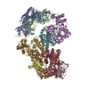
|
|---|---|
| 1 |
|
-Components
| #1: Protein | Mass: 32717.416 Da / Num. of mol.: 4 Source method: isolated from a genetically manipulated source Source: (gene. exp.) References: UniProt: A0A3W2RQG2, UniProt: P77398*PLUS, UDP-4-amino-4-deoxy-L-arabinose formyltransferase, UDP-glucuronic acid dehydrogenase (UDP-4-keto-hexauronic acid decarboxylating) #2: Protein | Mass: 40057.586 Da / Num. of mol.: 4 Source method: isolated from a genetically manipulated source Source: (gene. exp.) |
|---|
-Experimental details
-Experiment
| Experiment | Method: ELECTRON MICROSCOPY |
|---|---|
| EM experiment | Aggregation state: PARTICLE / 3D reconstruction method: single particle reconstruction |
- Sample preparation
| Component | Name: ArnA / Type: COMPLEX / Details: Full length ArnA from E.coli / Entity ID: all / Source: RECOMBINANT | ||||||||||||||||||||
|---|---|---|---|---|---|---|---|---|---|---|---|---|---|---|---|---|---|---|---|---|---|
| Molecular weight | Value: 0.44 MDa / Experimental value: YES | ||||||||||||||||||||
| Source (natural) | Organism: | ||||||||||||||||||||
| Source (recombinant) | Organism: | ||||||||||||||||||||
| Buffer solution | pH: 8 | ||||||||||||||||||||
| Buffer component |
| ||||||||||||||||||||
| Specimen | Conc.: 1.2 mg/ml / Embedding applied: NO / Shadowing applied: NO / Staining applied: NO / Vitrification applied: YES | ||||||||||||||||||||
| Vitrification | Cryogen name: ETHANE / Humidity: 100 % / Chamber temperature: 298 K |
- Electron microscopy imaging
| Experimental equipment |  Model: Titan Krios / Image courtesy: FEI Company |
|---|---|
| Microscopy | Model: FEI TITAN KRIOS |
| Electron gun | Electron source:  FIELD EMISSION GUN / Accelerating voltage: 300 kV / Illumination mode: SPOT SCAN FIELD EMISSION GUN / Accelerating voltage: 300 kV / Illumination mode: SPOT SCAN |
| Electron lens | Mode: BRIGHT FIELD / Cs: 2.7 mm |
| Image recording | Electron dose: 35 e/Å2 / Film or detector model: FEI FALCON II (4k x 4k) |
- Processing
| CTF correction | Type: PHASE FLIPPING AND AMPLITUDE CORRECTION |
|---|---|
| 3D reconstruction | Resolution: 7.8 Å / Resolution method: FSC 0.143 CUT-OFF / Num. of particles: 38127 / Symmetry type: POINT |
