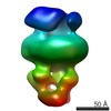
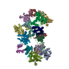
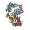
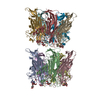
Journal: Front Mol Biosci / Year: 2017 Title: Electron Microscopy Structural Insights into CPAP Oligomeric Behavior: A Plausible Assembly Process of a Supramolecular Scaffold of the Centrosome. Authors: Ana L Alvarez-Cabrera / Sandra Delgado / David Gil-Carton / Gulnahar B Mortuza / Guillermo Montoya / Carlos O S Sorzano / Tang K Tang / Jose M Carazo / Abstract: Centrosomal P4.1-associated protein (CPAP) is a cell cycle regulated protein fundamental for centrosome assembly and centriole elongation. In humans, the region between residues 897-1338 of CPAP ...Centrosomal P4.1-associated protein (CPAP) is a cell cycle regulated protein fundamental for centrosome assembly and centriole elongation. In humans, the region between residues 897-1338 of CPAP mediates interactions with other proteins and includes a homodimerization domain. CPAP mutations cause primary autosomal recessive microcephaly and Seckel syndrome. Despite of the biological/clinical relevance of CPAP, its mechanistic behavior remains unclear and its C-terminus (the G-box/TCP domain) is the only part whose structure has been solved. This situation is perhaps due in part to the challenges that represent obtaining the protein in a soluble, homogeneous state for structural studies. Our work constitutes a systematic structural analysis on multiple oligomers of , using single-particle electron microscopy (EM) of negatively stained (NS) samples. Based on image classification into clearly different regular 3D maps (putatively corresponding to dimers and tetramers) and direct observation of individual images representing other complexes of CPAP (i.e., putative flexible monomers and higher-order multimers), we report a dynamic oligomeric behavior of this protein, where different homo-oligomers coexist in variable proportions. We propose that dimerization of the putative homodimer forms a putative tetramer which could be the structural unit for the scaffold that either tethers the pericentriolar material to centrioles or promotes procentriole elongation. A coarse fitting of atomic models into the NS 3D maps at resolutions around 20 Å is performed only to complement our experimental data, allowing us to hypothesize on the oligomeric composition of the different complexes. In this way, the current EM work represents an initial step toward the structural characterization of different oligomers of CPAP, suggesting further insights to understand how this protein works, contributing to the elucidation of control mechanisms for centriole biogenesis.
History
Deposition
Jul 13, 2016
-
Header (metadata) release
Sep 14, 2016
-
Map release
Sep 14, 2016
-
Update
Apr 18, 2018
-
Current status
Apr 18, 2018
Processing site: PDBe / Status: Released
-
Structure visualization
Movie
Surface view with section colored by density value
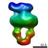
Entire : Putative homotetramer of human CPAP (residues 897-1338)
Entire
Name: Putative homotetramer of human CPAP (residues 897-1338)
Components
Complex: Putative homotetramer of human CPAP (residues 897-1338)
-
Supramolecule #1: Putative homotetramer of human CPAP (residues 897-1338)
Supramolecule
Name: Putative homotetramer of human CPAP (residues 897-1338) type: complex / ID: 1 / Parent: 0 Details: Human CPAP (residues 897-1338) expresed in E. coli and purified by Immobilized Metal Affinity Chromatography (IMAC) and Size Exclusion Chromatography (SEC)
Details: TCEP added to the buffer was prepared fresh
Staining
Type: NEGATIVE / Material: Uranyl Formate (0.75%)
Grid
Model: Quantifoil. Formvar/Carbon / Material: COPPER / Mesh: 400 / Support film - #0 - Film type ID: 1 / Support film - #0 - Material: CARBON / Support film - #0 - topology: CONTINUOUS / Support film - #1 - Film type ID: 2 / Support film - #1 - Material: FORMVAR / Support film - #1 - topology: CONTINUOUS / Pretreatment - Type: GLOW DISCHARGE
Details
This human CPAP construct includes residues 897-1338
-
Electron microscopy
Microscope
FEI TECNAI F20
Image recording
Film or detector model: FEI EAGLE (4k x 4k) / Number grids imaged: 1 / Average exposure time: 1.0 sec. / Average electron dose: 9.0 e/Å2
Electron beam
Acceleration voltage: 200 kV / Electron source: TUNGSTEN HAIRPIN
Electron optics
Illumination mode: SPOT SCAN / Imaging mode: BRIGHT FIELD / Cs: 2.26 mm
Experimental equipment
Model: Tecnai F20 / Image courtesy: FEI Company
+
Image processing
Particle selection
Number selected: 3048
CTF correction
Software - Name: Xmipp (ver. 3.1)
Startup model
Type of model: OTHER Details: RANSAC algorithm (Bioinformatics. 2014 Oct 15;30(20):2891-8)
Final reconstruction
Applied symmetry - Point group: C1 (asymmetric) / Resolution.type: BY AUTHOR / Resolution: 23.0 Å / Resolution method: FSC 0.143 CUT-OFF / Software - Name: Xmipp (ver. 3.1) / Software - details: Reconstruct Significant algorithm / Number images used: 3048
In the structure databanks used in Yorodumi, some data are registered as the other names, "COVID-19 virus" and "2019-nCoV". Here are the details of the virus and the list of structure data.
Jan 31, 2019. EMDB accession codes are about to change! (news from PDBe EMDB page)
EMDB accession codes are about to change! (news from PDBe EMDB page)
The allocation of 4 digits for EMDB accession codes will soon come to an end. Whilst these codes will remain in use, new EMDB accession codes will include an additional digit and will expand incrementally as the available range of codes is exhausted. The current 4-digit format prefixed with “EMD-” (i.e. EMD-XXXX) will advance to a 5-digit format (i.e. EMD-XXXXX), and so on. It is currently estimated that the 4-digit codes will be depleted around Spring 2019, at which point the 5-digit format will come into force.
The EM Navigator/Yorodumi systems omit the EMD- prefix.
Related info.:Q: What is EMD? / ID/Accession-code notation in Yorodumi/EM Navigator
Yorodumi is a browser for structure data from EMDB, PDB, SASBDB, etc.
This page is also the successor to EM Navigator detail page, and also detail information page/front-end page for Omokage search.
The word "yorodu" (or yorozu) is an old Japanese word meaning "ten thousand". "mi" (miru) is to see.
Related info.:EMDB / PDB / SASBDB / Comparison of 3 databanks / Yorodumi Search / Aug 31, 2016. New EM Navigator & Yorodumi / Yorodumi Papers / Jmol/JSmol / Function and homology information / Changes in new EM Navigator and Yorodumi
 Movie
Movie Controller


 Open data
 Basic information
 Map data
 Sample
 Homo sapiens (human)
 Authors
 Citation


 Structure visualization
 Movie viewer
 UCSF Chimera
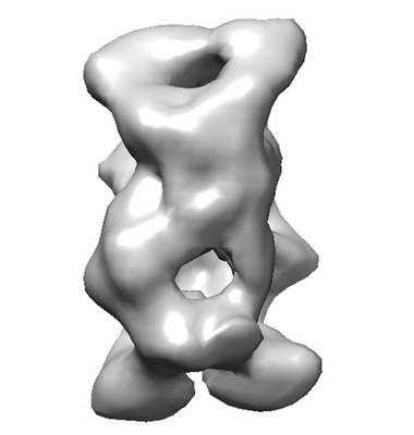
 Downloads & links
 emd_8288.png
 http://ftp.pdbj.org/pub/emdb/structures/EMD-8288
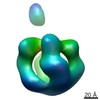
 Z (Sec.)
 Y (Row.)
 X (Col.)


 Sample components
 Processing
 Electron microscopy
