 Movie
Movie Controller

[English] 日本語
 Yorodumi
- PDB-6nt4: Cryo-EM structure of a human-cockroach hybrid Nav channel bound t... -
+ Open data

- Basic information
| Entry | Database: PDB / ID: 6nt4 | |||||||||||||||
|---|---|---|---|---|---|---|---|---|---|---|---|---|---|---|---|---|
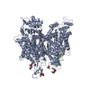
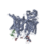
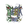
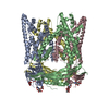
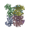
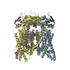
| Title | Cryo-EM structure of a human-cockroach hybrid Nav channel bound to alpha-scorpion toxin AaH2. | |||||||||||||||
 Components Components |
| |||||||||||||||
 Keywords Keywords | MEMBRANE PROTEIN / Sodium channel / scorpion toxin / electrical signaling / fast inactivation | |||||||||||||||
| Function / homology |  Function and homology information Function and homology informationdetection of mechanical stimulus involved in sensory perception / membrane depolarization during action potential / cardiac muscle cell action potential involved in contraction / voltage-gated sodium channel complex / sodium channel inhibitor activity / Interaction between L1 and Ankyrins / voltage-gated sodium channel activity / sodium ion transport / Phase 0 - rapid depolarisation / detection of temperature stimulus involved in sensory perception of pain ...detection of mechanical stimulus involved in sensory perception / membrane depolarization during action potential / cardiac muscle cell action potential involved in contraction / voltage-gated sodium channel complex / sodium channel inhibitor activity / Interaction between L1 and Ankyrins / voltage-gated sodium channel activity / sodium ion transport / Phase 0 - rapid depolarisation / detection of temperature stimulus involved in sensory perception of pain / behavioral response to pain / neuronal action potential / sodium ion transmembrane transport / sensory perception of pain / post-embryonic development / defense response / Sensory perception of sweet, bitter, and umami (glutamate) taste / response to toxic substance / circadian rhythm / toxin activity / inflammatory response / axon / extracellular region / plasma membrane Similarity search - Function | |||||||||||||||
| Biological species |  Periplaneta americana (American cockroach) Periplaneta americana (American cockroach) Homo sapiens (human)Androctonus australis (Sahara scorpion) Homo sapiens (human)Androctonus australis (Sahara scorpion) | |||||||||||||||
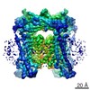
| Method | ELECTRON MICROSCOPY / single particle reconstruction / cryo EM / Resolution: 3.5 Å | |||||||||||||||
 Authors Authors | Clairfeuille, T. / Rohou, A. / Payandeh, J. | |||||||||||||||
 Citation Citation | Journal: Science / Year: 2019 Title: Structural basis of α-scorpion toxin action on Na channels. Authors: Thomas Clairfeuille / Alexander Cloake / Daniel T Infield / José P Llongueras / Christopher P Arthur / Zhong Rong Li / Yuwen Jian / Marie-France Martin-Eauclaire / Pierre E Bougis / Claudio ...Authors: Thomas Clairfeuille / Alexander Cloake / Daniel T Infield / José P Llongueras / Christopher P Arthur / Zhong Rong Li / Yuwen Jian / Marie-France Martin-Eauclaire / Pierre E Bougis / Claudio Ciferri / Christopher A Ahern / Frank Bosmans / David H Hackos / Alexis Rohou / Jian Payandeh /     Abstract: Fast inactivation of voltage-gated sodium (Na) channels is essential for electrical signaling, but its mechanism remains poorly understood. Here we determined the structures of a eukaryotic Na ...Fast inactivation of voltage-gated sodium (Na) channels is essential for electrical signaling, but its mechanism remains poorly understood. Here we determined the structures of a eukaryotic Na channel alone and in complex with a lethal α-scorpion toxin, AaH2, by electron microscopy, both at 3.5-angstrom resolution. AaH2 wedges into voltage-sensing domain IV (VSD4) to impede fast activation by trapping a deactivated state in which gating charge interactions bridge to the acidic intracellular carboxyl-terminal domain. In the absence of AaH2, the S4 helix of VSD4 undergoes a ~13-angstrom translation to unlatch the intracellular fast-inactivation gating machinery. Highlighting the polypharmacology of α-scorpion toxins, AaH2 also targets an unanticipated receptor site on VSD1 and a pore glycan adjacent to VSD4. Overall, this work provides key insights into fast inactivation, electromechanical coupling, and pathogenic mutations in Na channels. | |||||||||||||||
| History |
|
- Structure visualization
| Movie |
Movie viewer |
|---|---|
| Structure viewer | Molecule: MolmilJmol/JSmol |
- Downloads & links
-Download
| PDBx/mmCIF format | 6nt4.cif.gz | 278.9 KB | Display | PDBx/mmCIF format |
|---|---|---|---|---|
| PDB format | pdb6nt4.ent.gz | 225.6 KB | Display | PDB format |
| PDBx/mmJSON format | 6nt4.json.gz | Tree view | PDBx/mmJSON format | |
| Others |  Other downloads Other downloads |
-Validation report
| Summary document | 6nt4_validation.pdf.gz | 1.6 MB | Display | wwPDB validaton report |
|---|---|---|---|---|
| Full document | 6nt4_full_validation.pdf.gz | 1.6 MB | Display | |
| Data in XML | 6nt4_validation.xml.gz | 59.3 KB | Display | |
| Data in CIF | 6nt4_validation.cif.gz | 85.6 KB | Display | |
| Arichive directory | https://data.pdbj.org/pub/pdb/validation_reports/nt/6nt4ftp://data.pdbj.org/pub/pdb/validation_reports/nt/6nt4 | HTTPS FTP |
-Related structure data
| Related structure data | 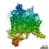 0501MC 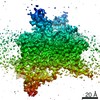 0500C  6nt3C C: citing same article ( M: map data used to model this data |
|---|---|
| Similar structure data |
-Links
 PDBj
PDBj


- Assembly
| Deposited unit | 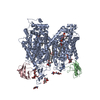
|
|---|---|
| 1 |
|
-Components
-Protein , 2 types, 3 molecules ABC
| #1: Protein | Mass: 178763.656 Da / Num. of mol.: 1 Source method: isolated from a genetically manipulated source Source: (gene. exp.) Periplaneta americana (American cockroach), (gene. exp.) Homo sapiens (human)Gene: SCN9A, NENA / Cell (production host): Expi293 / Production host: Homo sapiens (human) / References: UniProt: D0E0C2, UniProt: Q15858 |
|---|---|
| #2: Protein | Mass: 7260.175 Da / Num. of mol.: 2 / Source method: isolated from a natural source / Source: (natural) Androctonus australis (Sahara scorpion) / References: UniProt: P01484 |
-Sugars , 2 types, 6 molecules 
| #3: Polysaccharide | beta-D-mannopyranose-(1-3)-[beta-D-mannopyranose-(1-6)]beta-D-mannopyranose-(1-3)-2-acetamido-2- ...beta-D-mannopyranose-(1-3)-[beta-D-mannopyranose-(1-6)]beta-D-mannopyranose-(1-3)-2-acetamido-2-deoxy-beta-D-glucopyranose-(1-3)-2-acetamido-2-deoxy-beta-D-glucopyranose Source method: isolated from a genetically manipulated source |
|---|---|
| #4: Sugar | ChemComp-NAG /  Type: D-saccharide, beta linking / Mass: 221.208 Da / Num. of mol.: 5 Type: D-saccharide, beta linking / Mass: 221.208 Da / Num. of mol.: 5Source method: isolated from a genetically manipulated source Formula: C8H15NO6 |
-Non-polymers , 4 types, 8 molecules 


| #5: Chemical | ChemComp-76F / (  Mass: 742.018 Da / Num. of mol.: 5 / Source method: obtained synthetically / Formula: C41H76NO8P Mass: 742.018 Da / Num. of mol.: 5 / Source method: obtained synthetically / Formula: C41H76NO8P#6: Chemical | ChemComp-AJP / |  Mass: 1229.312 Da / Num. of mol.: 1 / Source method: obtained synthetically / Formula: C56H92O29 / Comment: detergent*YM Mass: 1229.312 Da / Num. of mol.: 1 / Source method: obtained synthetically / Formula: C56H92O29 / Comment: detergent*YM#7: Chemical | ChemComp-LHG / |  Mass: 722.970 Da / Num. of mol.: 1 / Source method: obtained synthetically / Formula: C38H75O10P / Comment: phospholipid*YM Mass: 722.970 Da / Num. of mol.: 1 / Source method: obtained synthetically / Formula: C38H75O10P / Comment: phospholipid*YM#8: Chemical | ChemComp-Y01 / |  Mass: 486.726 Da / Num. of mol.: 1 / Source method: obtained synthetically / Formula: C31H50O4 Mass: 486.726 Da / Num. of mol.: 1 / Source method: obtained synthetically / Formula: C31H50O4 |
|---|
-Experimental details
-Experiment
| Experiment | Method: ELECTRON MICROSCOPY |
|---|---|
| EM experiment | Aggregation state: PARTICLE / 3D reconstruction method: single particle reconstruction |
- Sample preparation
| Component | Name: NavPas-VSD4-AaH2 / Type: COMPLEX / Entity ID: #2 / Source: RECOMBINANT |
|---|---|
| Molecular weight | Value: 0.155 MDa / Experimental value: NO |
| Source (natural) | Organism: Periplaneta americana (American cockroach) |
| Source (recombinant) | Organism: Homo sapiens (human) / Cell: Expi293 |
| Buffer solution | pH: 7.5 |
| Specimen | Conc.: 1.5 mg/ml / Embedding applied: NO / Shadowing applied: NO / Staining applied: NO / Vitrification applied: YES |
| Specimen support | Details: Grids were plasma etched using the Solarus plasma cleaner (Gatan) in the hydrogen-oxygen setting. Grids were etched for 4 minutes on each side to remove burrs from hole edges. The grids were ...Details: Grids were plasma etched using the Solarus plasma cleaner (Gatan) in the hydrogen-oxygen setting. Grids were etched for 4 minutes on each side to remove burrs from hole edges. The grids were then coated on both sides with 5nm of Au/Pd which was plasma deposited using the Leica ACE600 (Leica). Grid material: COPPER / Grid mesh size: 200 divisions/in. / Grid type: C-flat-2/1 |
| Vitrification | Instrument: FEI VITROBOT MARK IV / Cryogen name: ETHANE / Humidity: 100 % / Chamber temperature: 277 K |
- Electron microscopy imaging
| Experimental equipment |  Model: Titan Krios / Image courtesy: FEI Company |
|---|---|
| Microscopy | Model: FEI TITAN KRIOS |
| Electron gun | Electron source:  FIELD EMISSION GUN / Accelerating voltage: 300 kV / Illumination mode: FLOOD BEAM FIELD EMISSION GUN / Accelerating voltage: 300 kV / Illumination mode: FLOOD BEAM |
| Electron lens | Mode: BRIGHT FIELD / Cs: 2.7 mm / Alignment procedure: ZEMLIN TABLEAU |
| Specimen holder | Cryogen: NITROGEN / Specimen holder model: FEI TITAN KRIOS AUTOGRID HOLDER |
| Image recording | Average exposure time: 10 sec. / Electron dose: 40 e/Å2 / Detector mode: COUNTING / Film or detector model: GATAN K2 SUMMIT (4k x 4k) / Num. of grids imaged: 1 / Num. of real images: 14886 |
| EM imaging optics | Energyfilter name: GIF Bioquantum / Energyfilter slit width: 20 eV |
| Image scans | Movie frames/image: 40 |
- Processing
| Software | Name: PHENIX / Version: 1.14_3260: / Classification: refinement | ||||||||||||||||||||||||
|---|---|---|---|---|---|---|---|---|---|---|---|---|---|---|---|---|---|---|---|---|---|---|---|---|---|
| EM software |
| ||||||||||||||||||||||||
| CTF correction | Type: PHASE FLIPPING AND AMPLITUDE CORRECTION | ||||||||||||||||||||||||
| Particle selection | Details: Automated picking using a soft-edged disc as a template yielded 842,024 candidate coordinates. 2D classification led to the selection of 504,252 particles for further analysis. | ||||||||||||||||||||||||
| Symmetry | Point symmetry: C1 (asymmetric) | ||||||||||||||||||||||||
| 3D reconstruction | Resolution: 3.5 Å / Resolution method: FSC 0.143 CUT-OFF / Num. of particles: 251714 / Algorithm: FOURIER SPACE Details: No information from spatial frequencies higher than 1/5A were ever used during refinement. Num. of class averages: 1 / Symmetry type: POINT |
