 Movie
Movie Controller


+ Open data

- Basic information
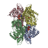
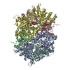
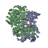
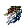
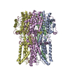
| Entry | Database: PDB / ID: 6l2t | ||||||
|---|---|---|---|---|---|---|---|
| Title | African swine fever virus major capsid protein p72 | ||||||
 Components Components | B646L,Major capsid protein | ||||||
 Keywords Keywords | VIRAL PROTEIN / African Swine Fever virus capsomer / Trisymmetron / NCLDV | ||||||
| Function / homology | Major capsid protein, C-terminal / Major capsid protein, C-terminal domain superfamily / Large eukaryotic DNA virus major capsid protein / Group II dsDNA virus coat/capsid protein / viral capsid / structural molecule activity / B646L Function and homology information Function and homology information | ||||||
| Biological species |   African swine fever virus African swine fever virus | ||||||
| Method | ELECTRON MICROSCOPY / single particle reconstruction / cryo EM / Resolution: 4.1 Å | ||||||
 Authors Authors | Wang, N. / Rao, Z. / Wang, X. | ||||||
 Citation Citation | Journal: Science / Year: 2019 Title: Architecture of African swine fever virus and implications for viral assembly. Authors: Nan Wang / Dongming Zhao / Jialing Wang / Yangling Zhang / Ming Wang / Yan Gao / Fang Li / Jingfei Wang / Zhigao Bu / Zihe Rao / Xiangxi Wang /  Abstract: African swine fever virus (ASFV) is a giant and complex DNA virus that causes a highly contagious and often lethal swine disease for which no vaccine is available. Using an optimized image ...African swine fever virus (ASFV) is a giant and complex DNA virus that causes a highly contagious and often lethal swine disease for which no vaccine is available. Using an optimized image reconstruction strategy, we solved the ASFV capsid structure up to 4.1 angstroms, which is built from 17,280 proteins, including one major (p72) and four minor (M1249L, p17, p49, and H240R) capsid proteins organized into pentasymmetrons and trisymmetrons. The atomic structure of the p72 protein informs putative conformational epitopes, distinguishing ASFV from other nucleocytoplasmic large DNA viruses. The minor capsid proteins form a complicated network below the outer capsid shell, stabilizing the capsid by holding adjacent capsomers together. Acting as core organizers, 100-nanometer-long M1249L proteins run along each edge of the trisymmetrons that bridge two neighboring pentasymmetrons and form extensive intermolecular networks with other capsid proteins, driving the formation of the capsid framework. These structural details unveil the basis of capsid stability and assembly, opening up new avenues for African swine fever vaccine development. | ||||||
| History |
|
- Structure visualization
| Movie |
Movie viewer |
|---|---|
| Structure viewer | Molecule: MolmilJmol/JSmol |
- Downloads & links
-Download
| PDBx/mmCIF format | 6l2t.cif.gz | 313.9 KB | Display | PDBx/mmCIF format |
|---|---|---|---|---|
| PDB format | pdb6l2t.ent.gz | 249.8 KB | Display | PDB format |
| PDBx/mmJSON format | 6l2t.json.gz | Tree view | PDBx/mmJSON format | |
| Others |  Other downloads Other downloads |
-Validation report
| Summary document | 6l2t_validation.pdf.gz | 778 KB | Display | wwPDB validaton report |
|---|---|---|---|---|
| Full document | 6l2t_full_validation.pdf.gz | 789.7 KB | Display | |
| Data in XML | 6l2t_validation.xml.gz | 51.3 KB | Display | |
| Data in CIF | 6l2t_validation.cif.gz | 77.2 KB | Display | |
| Arichive directory | https://data.pdbj.org/pub/pdb/validation_reports/l2/6l2tftp://data.pdbj.org/pub/pdb/validation_reports/l2/6l2t | HTTPS FTP |
-Related structure data
| Related structure data | 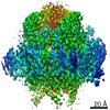 0814MC 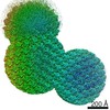 0810C 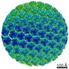 0811C 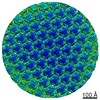 0812C 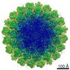 0813C 0815C M: map data used to model this data C: citing same article ( |
|---|---|
| Similar structure data |

-Links
 PDBj
PDBj

- Assembly
| Deposited unit | 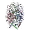
|
|---|---|
| 1 |
|
-Components
| #1: Protein | Mass: 73219.562 Da / Num. of mol.: 3 / Source method: isolated from a natural source / Source: (natural) African swine fever virus / References: UniProt: Q5IZK2*PLUSSequence details | Authors state that this intact virus sequence data were deposited in GenBank with the accession ...Authors state that this intact virus sequence data were deposited in GenBank with the accession numbers from MK333180 to MK333192. | |
|---|
-Experimental details
-Experiment
| Experiment | Method: ELECTRON MICROSCOPY |
|---|---|
| EM experiment | Aggregation state: PARTICLE / 3D reconstruction method: single particle reconstruction |
- Sample preparation
| Component | Name: African swine fever virus / Type: VIRUS / Entity ID: all / Source: NATURAL |
|---|---|
| Source (natural) | Organism: African swine fever virus |
| Details of virus | Empty: NO / Enveloped: YES / Isolate: SPECIES / Type: VIRION |
| Buffer solution | pH: 7 |
| Specimen | Embedding applied: NO / Shadowing applied: NO / Staining applied: NO / Vitrification applied: YES |
| Vitrification | Cryogen name: ETHANE |
- Electron microscopy imaging
| Experimental equipment |  Model: Titan Krios / Image courtesy: FEI Company |
|---|---|
| Microscopy | Model: FEI TITAN KRIOS |
| Electron gun | Electron source:  FIELD EMISSION GUN / Accelerating voltage: 300 kV / Illumination mode: FLOOD BEAM FIELD EMISSION GUN / Accelerating voltage: 300 kV / Illumination mode: FLOOD BEAM |
| Electron lens | Mode: BRIGHT FIELD |
| Image recording | Electron dose: 35 e/Å2 / Film or detector model: GATAN K2 QUANTUM (4k x 4k) |
- Processing
| Software | Name: PHENIX / Version: 1.17.1_3660: / Classification: refinement | ||||||||||||||||||||||||
|---|---|---|---|---|---|---|---|---|---|---|---|---|---|---|---|---|---|---|---|---|---|---|---|---|---|
| CTF correction | Type: NONE | ||||||||||||||||||||||||
| 3D reconstruction | Resolution: 4.1 Å / Resolution method: FSC 0.143 CUT-OFF / Num. of particles: 43811 / Symmetry type: POINT | ||||||||||||||||||||||||
| Refine LS restraints |
|
