Movie
Movie Controller


+ Open data

- Basic information
| Entry | Database: PDB / ID: 6ig9 | ||||||
|---|---|---|---|---|---|---|---|
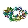
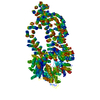
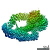
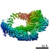
| Title | Tra1 subunit from Saccharomyces cerevisiae SAGA complex | ||||||
 Components Components | Transcription-associated protein 1 | ||||||
 Keywords Keywords | TRANSCRIPTION / Epigenetics / Acetyltransferase / Co-activator | ||||||
| Function / homology |  Function and homology information Function and homology informationTTT Hsp90 cochaperone complex / SLIK (SAGA-like) complex / SAGA complex / DNA repair-dependent chromatin remodeling / NuA4 histone acetyltransferase complex / Ub-specific processing proteases / DNA repair / regulation of transcription by RNA polymerase II / regulation of DNA-templated transcription / DNA-templated transcription ...TTT Hsp90 cochaperone complex / SLIK (SAGA-like) complex / SAGA complex / DNA repair-dependent chromatin remodeling / NuA4 histone acetyltransferase complex / Ub-specific processing proteases / DNA repair / regulation of transcription by RNA polymerase II / regulation of DNA-templated transcription / DNA-templated transcription / positive regulation of transcription by RNA polymerase II / nucleus Similarity search - Function | ||||||
| Biological species |  | ||||||
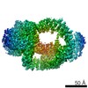
| Method | ELECTRON MICROSCOPY / single particle reconstruction / cryo EM / Resolution: 4.6 Å | ||||||
 Authors Authors | Zheng, X.D. / Liu, G.C. / Guan, H.P. / Li, H.T. | ||||||
 Citation Citation | Journal: Cell Discov / Year: 2019 Title: Architecture of SAGA complex. Authors: Gaochao Liu / Xiangdong Zheng / Haipeng Guan / Yong Cao / Hongyuan Qu / Junqing Kang / Xiangle Ren / Jianlin Lei / Meng-Qiu Dong / Xueming Li / Haitao Li /  | ||||||
| History |
|
- Structure visualization
| Movie |
Movie viewer |
|---|---|
| Structure viewer | Molecule: MolmilJmol/JSmol |
- Downloads & links
-Download
| PDBx/mmCIF format | 6ig9.cif.gz | 492.5 KB | Display | PDBx/mmCIF format |
|---|---|---|---|---|
| PDB format | pdb6ig9.ent.gz | 333.5 KB | Display | PDB format |
| PDBx/mmJSON format | 6ig9.json.gz | Tree view | PDBx/mmJSON format | |
| Others |  Other downloads Other downloads |
-Validation report
| Arichive directory | https://data.pdbj.org/pub/pdb/validation_reports/ig/6ig9ftp://data.pdbj.org/pub/pdb/validation_reports/ig/6ig9 | HTTPS FTP |
|---|
-Related structure data
| Related structure data | 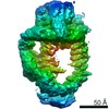 9664MC 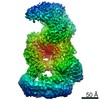 9663C M: map data used to model this data C: citing same article ( |
|---|---|
| Similar structure data |


-Links
 PDBj
PDBj


- Assembly
| Deposited unit | 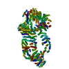
|
|---|---|
| 1 |
|
-Components
| #1: Protein | Mass: 433677.281 Da / Num. of mol.: 1 / Source method: isolated from a natural source / Source: (natural) |
|---|
-Experimental details
-Experiment
| Experiment | Method: ELECTRON MICROSCOPY |
|---|---|
| EM experiment | Aggregation state: PARTICLE / 3D reconstruction method: single particle reconstruction |
- Sample preparation
| Component | Name: Tra1 / ScTra1 / Type: COMPLEX Details: One of the major part from SAGA (Spt-Ada-Gcn5-Acetyltransferase) complex Entity ID: all / Source: NATURAL | ||||||||||||
|---|---|---|---|---|---|---|---|---|---|---|---|---|---|
| Molecular weight | Value: 1.8 MDa / Experimental value: NO | ||||||||||||
| Source (natural) | Organism: | ||||||||||||
| Buffer solution | pH: 8.5 / Details: 20mM HEPES pH 8.5 150mM Nacl | ||||||||||||
| Buffer component |
| ||||||||||||
| Specimen | Conc.: 0.2 mg/ml / Embedding applied: NO / Shadowing applied: NO / Staining applied: NO / Vitrification applied: YES Details: We extracted it from the overall SAGA complex structure. | ||||||||||||
| Specimen support | Details: The grid was coated with a home-made thin continuous carbon film. Grid material: COPPER / Grid mesh size: 400 divisions/in. / Grid type: Quantifoil R2/2 | ||||||||||||
| Vitrification | Instrument: FEI VITROBOT MARK IV / Cryogen name: ETHANE / Humidity: 95 % / Chamber temperature: 281 K Details: Blot for 4 seconds and wait for 30 seconds before plunging |
- Electron microscopy imaging
| Experimental equipment |  Model: Titan Krios / Image courtesy: FEI Company |
|---|---|
| Microscopy | Model: FEI TITAN KRIOS |
| Electron gun | Electron source:  FIELD EMISSION GUN / Accelerating voltage: 300 kV / Illumination mode: FLOOD BEAM FIELD EMISSION GUN / Accelerating voltage: 300 kV / Illumination mode: FLOOD BEAM |
| Electron lens | Mode: BRIGHT FIELD / Nominal magnification: 81000 X / Calibrated magnification: 81000 X / Nominal defocus max: 3000 nm / Nominal defocus min: 1200 nm / Calibrated defocus min: 1200 nm / Calibrated defocus max: 3000 nm / Cs: 0.1 mm / C2 aperture diameter: 70 µm / Alignment procedure: COMA FREE |
| Specimen holder | Cryogen: NITROGEN / Specimen holder model: FEI TITAN KRIOS AUTOGRID HOLDER / Temperature (max): 70 K / Temperature (min): 70 K |
| Image recording | Average exposure time: 0.25 sec. / Electron dose: 5.6 e/Å2 / Detector mode: SUPER-RESOLUTION / Film or detector model: GATAN K2 SUMMIT (4k x 4k) / Num. of real images: 8526 |
| EM imaging optics | Spherical aberration corrector: With a Cs-corrector |
| Image scans | Sampling size: 5 µm / Width: 3838 / Height: 3710 / Movie frames/image: 32 / Used frames/image: 3-30 |
- Processing
| Software | Name: PHENIX / Version: dev_3290: / Classification: refinement | |||||||||||||||||||||||||||||||||||||||||||||
|---|---|---|---|---|---|---|---|---|---|---|---|---|---|---|---|---|---|---|---|---|---|---|---|---|---|---|---|---|---|---|---|---|---|---|---|---|---|---|---|---|---|---|---|---|---|---|
| EM software |
| |||||||||||||||||||||||||||||||||||||||||||||
| CTF correction | Details: GPU accelerated / Type: NONE | |||||||||||||||||||||||||||||||||||||||||||||
| Particle selection | Num. of particles selected: 1350000 / Details: GPU accelerated | |||||||||||||||||||||||||||||||||||||||||||||
| Symmetry | Point symmetry: C1 (asymmetric) | |||||||||||||||||||||||||||||||||||||||||||||
| 3D reconstruction | Resolution: 4.6 Å / Resolution method: FSC 0.143 CUT-OFF / Num. of particles: 176464 / Algorithm: FOURIER SPACE / Details: GPU accelerated / Num. of class averages: 1 / Symmetry type: POINT | |||||||||||||||||||||||||||||||||||||||||||||
| Atomic model building | Protocol: AB INITIO MODEL / Space: REAL | |||||||||||||||||||||||||||||||||||||||||||||
| Atomic model building | PDB-ID: 5OJS Accession code: 5OJS / Source name: PDB / Type: experimental model | |||||||||||||||||||||||||||||||||||||||||||||
| Refine LS restraints |
|