 Movie
Movie Controller


+ Open data

- Basic information
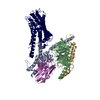
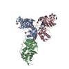
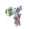
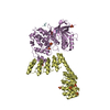
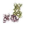
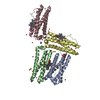
| Entry | Database: PDB / ID: 6ddf | ||||||||||||||||||
|---|---|---|---|---|---|---|---|---|---|---|---|---|---|---|---|---|---|---|---|
| Title | Mu Opioid Receptor-Gi Protein Complex | ||||||||||||||||||
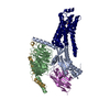
 Components Components |
| ||||||||||||||||||
 Keywords Keywords | MEMBRANE PROTEIN / Complex / Transmembrane | ||||||||||||||||||
| Function / homology |  Function and homology information Function and homology informationOpioid Signalling / spine apparatus / positive regulation of appetite / sperm ejaculation / Peptide ligand-binding receptors / G-protein activation / beta-endorphin receptor activity / morphine receptor activity / negative regulation of Wnt protein secretion / adenylate cyclase-inhibiting opioid receptor signaling pathway ...Opioid Signalling / spine apparatus / positive regulation of appetite / sperm ejaculation / Peptide ligand-binding receptors / G-protein activation / beta-endorphin receptor activity / morphine receptor activity / negative regulation of Wnt protein secretion / adenylate cyclase-inhibiting opioid receptor signaling pathway / negative regulation of luteinizing hormone secretion / G protein-coupled opioid receptor activity / regulation of cellular response to stress / G alpha (i) signalling events / G protein-coupled opioid receptor signaling pathway / negative regulation of adenylate cyclase-activating G protein-coupled receptor signaling pathway / negative regulation of nitric oxide biosynthetic process / adenylate cyclase-inhibiting G protein-coupled acetylcholine receptor signaling pathway / behavioral response to ethanol / regulation of NMDA receptor activity / neuropeptide binding / positive regulation of neurogenesis / eating behavior / negative regulation of cytosolic calcium ion concentration / transmission of nerve impulse / G-protein alpha-subunit binding / social behavior / voltage-gated calcium channel activity / adenylate cyclase inhibitor activity / positive regulation of protein localization to cell cortex / T cell migration / positive regulation of relaxation of smooth muscle / Adenylate cyclase inhibitory pathway / D2 dopamine receptor binding / T-tubule / sensory perception of pain / adenylate cyclase-inhibiting serotonin receptor signaling pathway / G protein-coupled serotonin receptor binding / positive regulation of gluconeogenesis / dendrite cytoplasm / dendrite membrane / presynaptic modulation of chemical synaptic transmission / cellular response to forskolin / regulation of mitotic spindle organization / chemokine-mediated signaling pathway / sarcoplasmic reticulum / Regulation of insulin secretion / neuropeptide signaling pathway / response to prostaglandin E / excitatory postsynaptic potential / locomotory behavior / positive regulation of cholesterol biosynthetic process / sarcolemma / negative regulation of insulin secretion / G protein-coupled receptor binding / response to peptide hormone / GABA-ergic synapse / G protein-coupled receptor activity / centriolar satellite / G-protein beta/gamma-subunit complex binding / adenylate cyclase-modulating G protein-coupled receptor signaling pathway / adenylate cyclase-inhibiting G protein-coupled receptor signaling pathway / Olfactory Signaling Pathway / Activation of the phototransduction cascade / G protein-coupled acetylcholine receptor signaling pathway / G beta:gamma signalling through PLC beta / Presynaptic function of Kainate receptors / Thromboxane signalling through TP receptor / Activation of G protein gated Potassium channels / Inhibition of voltage gated Ca2+ channels via Gbeta/gamma subunits / G-protein activation / Glucagon signaling in metabolic regulation / Prostacyclin signalling through prostacyclin receptor / G beta:gamma signalling through CDC42 / Synthesis, secretion, and inactivation of Glucagon-like Peptide-1 (GLP-1) / G beta:gamma signalling through BTK / photoreceptor disc membrane / ADP signalling through P2Y purinoceptor 12 / Glucagon-type ligand receptors / GDP binding / Sensory perception of sweet, bitter, and umami (glutamate) taste / Adrenaline,noradrenaline inhibits insulin secretion / Vasopressin regulates renal water homeostasis via Aquaporins / Glucagon-like Peptide-1 (GLP1) regulates insulin secretion / G alpha (z) signalling events / cellular response to catecholamine stimulus / ADP signalling through P2Y purinoceptor 1 / ADORA2B mediated anti-inflammatory cytokines production / G beta:gamma signalling through PI3Kgamma / adenylate cyclase-activating dopamine receptor signaling pathway / Cooperation of PDCL (PhLP1) and TRiC/CCT in G-protein beta folding / GPER1 signaling / cellular response to prostaglandin E stimulus / heterotrimeric G-protein complex / G alpha (12/13) signalling events / G-protein beta-subunit binding / Inactivation, recovery and regulation of the phototransduction cascade / extracellular vesicle / sensory perception of taste / sperm principal piece Similarity search - Function | ||||||||||||||||||
| Biological species |  Homo sapiens (human) Homo sapiens (human) | ||||||||||||||||||
| Method | ELECTRON MICROSCOPY / single particle reconstruction / cryo EM / Resolution: 3.5 Å | ||||||||||||||||||
 Authors Authors | Koehl, A. / Hu, H. / Maeda, S. / Manglik, A. / Kobilka, B.K. / Skiniotis, G. / Weis, W.I. | ||||||||||||||||||
| Funding support |  United States, United States,  Switzerland, 5items Switzerland, 5items
| ||||||||||||||||||
 Citation Citation | Journal: Nature / Year: 2018 Title: Structure of the µ-opioid receptor-G protein complex. Authors: Antoine Koehl / Hongli Hu / Shoji Maeda / Yan Zhang / Qianhui Qu / Joseph M Paggi / Naomi R Latorraca / Daniel Hilger / Roger Dawson / Hugues Matile / Gebhard F X Schertler / Sebastien ...Authors: Antoine Koehl / Hongli Hu / Shoji Maeda / Yan Zhang / Qianhui Qu / Joseph M Paggi / Naomi R Latorraca / Daniel Hilger / Roger Dawson / Hugues Matile / Gebhard F X Schertler / Sebastien Granier / William I Weis / Ron O Dror / Aashish Manglik / Georgios Skiniotis / Brian K Kobilka /  Abstract: The μ-opioid receptor (μOR) is a G-protein-coupled receptor (GPCR) and the target of most clinically and recreationally used opioids. The induced positive effects of analgesia and euphoria are ...The μ-opioid receptor (μOR) is a G-protein-coupled receptor (GPCR) and the target of most clinically and recreationally used opioids. The induced positive effects of analgesia and euphoria are mediated by μOR signalling through the adenylyl cyclase-inhibiting heterotrimeric G protein G. Here we present the 3.5 Å resolution cryo-electron microscopy structure of the μOR bound to the agonist peptide DAMGO and nucleotide-free G. DAMGO occupies the morphinan ligand pocket, with its N terminus interacting with conserved receptor residues and its C terminus engaging regions important for opioid-ligand selectivity. Comparison of the μOR-G complex to previously determined structures of other GPCRs bound to the stimulatory G protein G reveals differences in the position of transmembrane receptor helix 6 and in the interactions between the G protein α-subunit and the receptor core. Together, these results shed light on the structural features that contribute to the G protein-coupling specificity of the µOR. | ||||||||||||||||||
| History |
|
- Structure visualization
| Movie |
Movie viewer |
|---|---|
| Structure viewer | Molecule: MolmilJmol/JSmol |
- Downloads & links
-Download
| PDBx/mmCIF format | 6ddf.cif.gz | 172.1 KB | Display | PDBx/mmCIF format |
|---|---|---|---|---|
| PDB format | pdb6ddf.ent.gz | 128.4 KB | Display | PDB format |
| PDBx/mmJSON format | 6ddf.json.gz | Tree view | PDBx/mmJSON format | |
| Others |  Other downloads Other downloads |
-Validation report
| Arichive directory | https://data.pdbj.org/pub/pdb/validation_reports/dd/6ddfftp://data.pdbj.org/pub/pdb/validation_reports/dd/6ddf | HTTPS FTP |
|---|
-Related structure data
| Related structure data |  7869MC 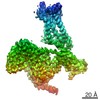 7868C 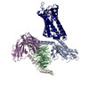 6ddeC C: citing same article ( M: map data used to model this data |
|---|---|
| Similar structure data |

-Links
 PDBj
PDBj


- Assembly
| Deposited unit | 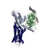
|
|---|---|
| 1 |
|
-Components
| #1: Protein | Mass: 40415.031 Da / Num. of mol.: 1 Source method: isolated from a genetically manipulated source Source: (gene. exp.) Homo sapiens (human) / Gene: GNAI1 / Plasmid: pVL1392 / Production host:  Trichoplusia ni (cabbage looper) / Strain (production host): High Five / References: UniProt: P63096 Trichoplusia ni (cabbage looper) / Strain (production host): High Five / References: UniProt: P63096 |
|---|---|
| #2: Protein | Mass: 37671.102 Da / Num. of mol.: 1 Source method: isolated from a genetically manipulated source Source: (gene. exp.) Homo sapiens (human) / Gene: GNB1 / Plasmid: pVL1392 / Production host: Trichoplusia ni (cabbage looper) / Strain (production host): High Five / References: UniProt: P62873 |
| #3: Protein | Mass: 7861.143 Da / Num. of mol.: 1 Source method: isolated from a genetically manipulated source Source: (gene. exp.) Homo sapiens (human) / Gene: GNG2 / Plasmid: pVL1392 / Production host: Trichoplusia ni (cabbage looper) / Strain (production host): High Five / References: UniProt: P59768 |
| #4: Protein | Mass: 39995.105 Da / Num. of mol.: 1 Source method: isolated from a genetically manipulated source Source: (gene. exp.)  Spodoptera frugiperda (fall armyworm) / Strain (production host): Sf9 / References: UniProt: P42866 Spodoptera frugiperda (fall armyworm) / Strain (production host): Sf9 / References: UniProt: P42866 |
| #5: Protein/peptide | 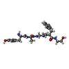  Type: Peptide-like / Mass: 513.587 Da / Num. of mol.: 1 / Source method: obtained synthetically / Details: analogue of enkephalin / Source: (synth.) Homo sapiens (human) / References: DAMGO Type: Peptide-like / Mass: 513.587 Da / Num. of mol.: 1 / Source method: obtained synthetically / Details: analogue of enkephalin / Source: (synth.) Homo sapiens (human) / References: DAMGO |
| Has protein modification | Y |
-Experimental details
-Experiment
| Experiment | Method: ELECTRON MICROSCOPY |
|---|---|
| EM experiment | Aggregation state: PARTICLE / 3D reconstruction method: single particle reconstruction |
- Sample preparation
| Component | Name: Ternary complex of DAMGO-activated Mu-type opioid receptor with heterotrimeric Gi Type: COMPLEX Details: Signaling complex formed by incubation of DAMGO-bound Mu-type opioid receptor and heterotrimeric Gi. Excess GDP removed by addition of apyrase. Entity ID: all / Source: RECOMBINANT | ||||||||||||||||||||||||||||||||||||||||
|---|---|---|---|---|---|---|---|---|---|---|---|---|---|---|---|---|---|---|---|---|---|---|---|---|---|---|---|---|---|---|---|---|---|---|---|---|---|---|---|---|---|
| Molecular weight | Value: 0.12 MDa / Experimental value: NO | ||||||||||||||||||||||||||||||||||||||||
| Source (natural) | Organism: Homo sapiens (human) | ||||||||||||||||||||||||||||||||||||||||
| Source (recombinant) | Organism: Spodoptera frugiperda (fall armyworm) | ||||||||||||||||||||||||||||||||||||||||
| Buffer solution | pH: 7.5 Details: Solutions were made fresh. After buffer reconstitution, all buffers were filtered with 0.22um filter. | ||||||||||||||||||||||||||||||||||||||||
| Buffer component |
| ||||||||||||||||||||||||||||||||||||||||
| Specimen | Conc.: 7 mg/ml / Embedding applied: NO / Shadowing applied: NO / Staining applied: NO / Vitrification applied: YES Details: This sample was monodisperse by negative stain and Cryo-EM analysis. | ||||||||||||||||||||||||||||||||||||||||
| Specimen support | Grid material: GOLD / Grid mesh size: 200 divisions/in. / Grid type: Quantifoil R1.2/1.3 | ||||||||||||||||||||||||||||||||||||||||
| Vitrification | Instrument: FEI VITROBOT MARK IV / Cryogen name: ETHANE / Humidity: 100 % / Chamber temperature: 293 K / Details: blot 1 second before plunging |
- Electron microscopy imaging
| Experimental equipment |  Model: Titan Krios / Image courtesy: FEI Company |
|---|---|
| Microscopy | Model: FEI TITAN KRIOS |
| Electron gun | Electron source:  FIELD EMISSION GUN / Accelerating voltage: 300 kV / Illumination mode: FLOOD BEAM FIELD EMISSION GUN / Accelerating voltage: 300 kV / Illumination mode: FLOOD BEAM |
| Electron lens | Mode: BRIGHT FIELD / Calibrated magnification: 48076 X / Nominal defocus min: 800 nm / Cs: 2.7 mm |
| Specimen holder | Cryogen: NITROGEN / Specimen holder model: FEI TITAN KRIOS AUTOGRID HOLDER |
| Image recording | Average exposure time: 8 sec. / Electron dose: 37 e/Å2 / Detector mode: COUNTING / Film or detector model: GATAN K2 SUMMIT (4k x 4k) / Num. of real images: 2642 Details: Images were collected in movie-mode at 10 frames per second. |
| Image scans | Movie frames/image: 80 |
- Processing
| EM software |
| ||||||||||||||||
|---|---|---|---|---|---|---|---|---|---|---|---|---|---|---|---|---|---|
| CTF correction | Type: PHASE FLIPPING ONLY | ||||||||||||||||
| Particle selection | Num. of particles selected: 893426 | ||||||||||||||||
| 3D reconstruction | Resolution: 3.5 Å / Resolution method: FSC 0.143 CUT-OFF / Num. of particles: 359406 / Symmetry type: POINT |
