 Movie
Movie Controller

[English] 日本語
 Yorodumi
- PDB-6dbj: Cryo-EM structure of RAG in complex with 12-RSS and 23-RSS nicked... -
+ Open data

- Basic information
| Entry | Database: PDB / ID: 6dbj | ||||||
|---|---|---|---|---|---|---|---|
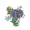
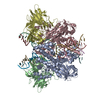
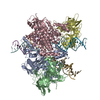
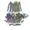
| Title | Cryo-EM structure of RAG in complex with 12-RSS and 23-RSS nicked DNA intermediates | ||||||
 Components Components |
| ||||||
 Keywords Keywords | RECOMBINATION/DNA / Synaptic RAG complex / V(D)J recombination / RSS / Paired complex / RECOMBINATION-DNA complex | ||||||
| Function / homology |  Function and homology information Function and homology informationsomatic diversification of immune receptors via germline recombination within a single locus / hematopoietic or lymphoid organ development / protein-DNA complex assembly / DNA recombinase complex / endodeoxyribonuclease complex / lymphocyte differentiation / pre-B cell allelic exclusion / immunoglobulin V(D)J recombination / V(D)J recombination / phosphatidylinositol-3,4-bisphosphate binding ...somatic diversification of immune receptors via germline recombination within a single locus / hematopoietic or lymphoid organ development / protein-DNA complex assembly / DNA recombinase complex / endodeoxyribonuclease complex / lymphocyte differentiation / pre-B cell allelic exclusion / immunoglobulin V(D)J recombination / V(D)J recombination / phosphatidylinositol-3,4-bisphosphate binding / histone H3K4me3 reader activity / phosphatidylinositol-3,5-bisphosphate binding / detection of maltose stimulus / maltose transport complex / phosphatidylinositol-3,4,5-trisphosphate binding / carbohydrate transport / T cell differentiation / carbohydrate transmembrane transporter activity / maltose binding / maltose transport / maltodextrin transmembrane transport / ATP-binding cassette (ABC) transporter complex, substrate-binding subunit-containing / phosphatidylinositol-4,5-bisphosphate binding / phosphatidylinositol binding / ATP-binding cassette (ABC) transporter complex / B cell differentiation / thymus development / cell chemotaxis / RING-type E3 ubiquitin transferase / ubiquitin-protein transferase activity / T cell differentiation in thymus / ubiquitin protein ligase activity / outer membrane-bounded periplasmic space / chromatin organization / endonuclease activity / histone binding / sequence-specific DNA binding / Hydrolases; Acting on ester bonds / adaptive immune response / periplasmic space / hydrolase activity / chromatin binding / DNA damage response / magnesium ion binding / protein homodimerization activity / zinc ion binding / membrane / metal ion binding / nucleus Similarity search - Function | ||||||
| Biological species |   | ||||||
| Method | ELECTRON MICROSCOPY / single particle reconstruction / cryo EM / Resolution: 3 Å | ||||||
 Authors Authors | Wu, H. / Liao, M. / Ru, H. / Mi, W. | ||||||
| Funding support |  United States, 1items United States, 1items
| ||||||
 Citation Citation | Journal: Nat Struct Mol Biol / Year: 2018 Title: DNA melting initiates the RAG catalytic pathway. Authors: Heng Ru / Wei Mi / Pengfei Zhang / Frederick W Alt / David G Schatz / Maofu Liao / Hao Wu / Abstract: The mechanism for initiating DNA cleavage by DDE-family enzymes, including the RAG endonuclease, which initiates V(D)J recombination, is not well understood. Here we report six cryo-EM structures of ...The mechanism for initiating DNA cleavage by DDE-family enzymes, including the RAG endonuclease, which initiates V(D)J recombination, is not well understood. Here we report six cryo-EM structures of zebrafish RAG in complex with one or two intact recombination signal sequences (RSSs), at up to 3.9-Å resolution. Unexpectedly, these structures reveal DNA melting at the heptamer of the RSSs, thus resulting in a corkscrew-like rotation of coding-flank DNA and the positioning of the scissile phosphate in the active site. Substrate binding is associated with dimer opening and a piston-like movement in RAG1, first outward to accommodate unmelted DNA and then inward to wedge melted DNA. These precleavage complexes show limited base-specific contacts of RAG at the conserved terminal CAC/GTG sequence of the heptamer, thus suggesting conservation based on a propensity to unwind. CA and TG overwhelmingly dominate terminal sequences in transposons and retrotransposons, thereby implicating a universal mechanism for DNA melting during the initiation of retroviral integration and DNA transposition. | ||||||
| History |
|
- Structure visualization
| Movie |
Movie viewer |
|---|---|
| Structure viewer | Molecule: MolmilJmol/JSmol |
- Downloads & links
-Download
| PDBx/mmCIF format | 6dbj.cif.gz | 474.1 KB | Display | PDBx/mmCIF format |
|---|---|---|---|---|
| PDB format | pdb6dbj.ent.gz | 353.3 KB | Display | PDB format |
| PDBx/mmJSON format | 6dbj.json.gz | Tree view | PDBx/mmJSON format | |
| Others |  Other downloads Other downloads |
-Validation report
| Arichive directory | https://data.pdbj.org/pub/pdb/validation_reports/db/6dbjftp://data.pdbj.org/pub/pdb/validation_reports/db/6dbj | HTTPS FTP |
|---|
-Related structure data
| Related structure data | 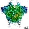 7844MC 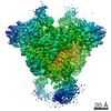 7843C 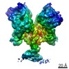 7845C 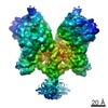 7846C 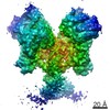 7847C 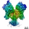 7848C 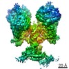 7849C 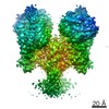 7850C 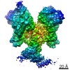 7851C 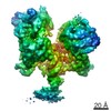 7852C 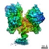 7853C  6dbiC 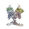 6dblC 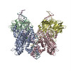 6dboC 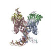 6dbqC 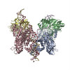 6dbrC 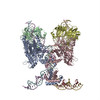 6dbtC 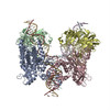 6dbuC 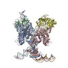 6dbvC 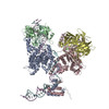 6dbwC 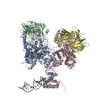 6dbxC C: citing same article ( M: map data used to model this data |
|---|---|
| Similar structure data |


-Links
 PDBj
PDBj


- Assembly
| Deposited unit | 
|
|---|---|
| 1 |
|
-Components
-Recombination activating gene ... , 2 types, 4 molecules ACBD
| #1: Protein | Mass: 131160.047 Da / Num. of mol.: 2 Source method: isolated from a genetically manipulated source Source: (gene. exp.) Strain: K12 / Gene: malE, b4034, JW3994, rag1 / Production host:   Spodoptera frugiperda (fall armyworm) Spodoptera frugiperda (fall armyworm)References: UniProt: P0AEX9, UniProt: O13033, RING-type E3 ubiquitin transferase #2: Protein | Mass: 59435.930 Da / Num. of mol.: 2 Source method: isolated from a genetically manipulated source Source: (gene. exp.) Spodoptera frugiperda (fall armyworm) / References: UniProt: Q1RLW7, UniProt: O13034*PLUS |
|---|
-DNA chain , 3 types, 6 molecules EHFGIJ
| #3: DNA chain | Mass: 4562.988 Da / Num. of mol.: 2 / Source method: obtained synthetically / Source: (synth.) #4: DNA chain | Mass: 9577.172 Da / Num. of mol.: 2 / Source method: obtained synthetically / Source: (synth.) #5: DNA chain | Mass: 4880.164 Da / Num. of mol.: 2 / Source method: obtained synthetically / Source: (synth.) |
|---|
-Non-polymers , 2 types, 6 molecules 

| #6: Chemical |  Mass: 65.409 Da / Num. of mol.: 2 / Source method: obtained synthetically / Formula: Zn Mass: 65.409 Da / Num. of mol.: 2 / Source method: obtained synthetically / Formula: Zn#7: Chemical | ChemComp-CA /  Mass: 40.078 Da / Num. of mol.: 4 / Source method: obtained synthetically / Formula: Ca Mass: 40.078 Da / Num. of mol.: 4 / Source method: obtained synthetically / Formula: Ca |
|---|
-Experimental details
-Experiment
| Experiment | Method: ELECTRON MICROSCOPY |
|---|---|
| EM experiment | Aggregation state: PARTICLE / 3D reconstruction method: single particle reconstruction |
- Sample preparation
| Component | Name: RAG in complex with 12-RSS and 23-RSS nicked DNA intermediates Type: COMPLEX / Entity ID: #1-#5 / Source: RECOMBINANT | |||||||||||||||||||||||||
|---|---|---|---|---|---|---|---|---|---|---|---|---|---|---|---|---|---|---|---|---|---|---|---|---|---|---|
| Source (natural) | Organism: | |||||||||||||||||||||||||
| Source (recombinant) | Organism: Spodoptera frugiperda (fall armyworm) | |||||||||||||||||||||||||
| Buffer solution | pH: 7.5 Details: Solutions were made fresh from concentrated to avoid microbial contamination. | |||||||||||||||||||||||||
| Buffer component |
| |||||||||||||||||||||||||
| Specimen | Embedding applied: NO / Shadowing applied: NO / Staining applied: NO / Vitrification applied: YES / Details: This sample was monodisperse. | |||||||||||||||||||||||||
| Vitrification | Cryogen name: ETHANE |
- Electron microscopy imaging
| Experimental equipment |  Model: Titan Krios / Image courtesy: FEI Company |
|---|---|
| Microscopy | Model: FEI TITAN KRIOS |
| Electron gun | Electron source:  FIELD EMISSION GUN / Accelerating voltage: 300 kV / Illumination mode: FLOOD BEAM FIELD EMISSION GUN / Accelerating voltage: 300 kV / Illumination mode: FLOOD BEAM |
| Electron lens | Mode: BRIGHT FIELD |
| Image recording | Electron dose: 40 e/Å2 / Film or detector model: GATAN K2 SUMMIT (4k x 4k) |
- Processing
| Software | Name: PHENIX / Version: (1.13_2998: ???) / Classification: refinement | ||||||||||||||||||||||||
|---|---|---|---|---|---|---|---|---|---|---|---|---|---|---|---|---|---|---|---|---|---|---|---|---|---|
| CTF correction | Type: PHASE FLIPPING AND AMPLITUDE CORRECTION | ||||||||||||||||||||||||
| Symmetry | Point symmetry: C2 (2 fold cyclic) | ||||||||||||||||||||||||
| 3D reconstruction | Resolution: 3 Å / Resolution method: FSC 0.143 CUT-OFF / Num. of particles: 196652 / Symmetry type: POINT | ||||||||||||||||||||||||
| Refinement | Resolution: 3→3 Å / SU ML: 0.79 / σ(F): 0.03 / Phase error: 46.56 / Stereochemistry target values: MLHL
| ||||||||||||||||||||||||
| Solvent computation | Shrinkage radii: 0.9 Å / VDW probe radii: 1.11 Å / Solvent model: FLAT BULK SOLVENT MODEL | ||||||||||||||||||||||||
| Refine LS restraints |
|
