 Movie
Movie Controller

[English] 日本語
 Yorodumi
- PDB-6ci1: The Structure of Full-Length Kv Beta 2.1 Determined by Cryogenic ... -
+ Open data

- Basic information
| Entry | Database: PDB / ID: 6ci1 | |||||||||
|---|---|---|---|---|---|---|---|---|---|---|
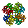
| Title | The Structure of Full-Length Kv Beta 2.1 Determined by Cryogenic Electron Microscopy | |||||||||
 Components Components | Voltage-gated potassium channel subunit beta-2 | |||||||||
 Keywords Keywords | OXIDOREDUCTASE / Kv Channel / Regulator / Complex | |||||||||
| Function / homology |  Function and homology information Function and homology informationpinceau fiber / methylglyoxal reductase (NADPH) (acetol producing) activity / Voltage gated Potassium channels / potassium channel complex / regulation of protein localization to cell surface / : / axon initial segment / juxtaparanode region of axon / Oxidoreductases; Acting on the CH-OH group of donors; With NAD+ or NADP+ as acceptor / myoblast differentiation ...pinceau fiber / methylglyoxal reductase (NADPH) (acetol producing) activity / Voltage gated Potassium channels / potassium channel complex / regulation of protein localization to cell surface / : / axon initial segment / juxtaparanode region of axon / Oxidoreductases; Acting on the CH-OH group of donors; With NAD+ or NADP+ as acceptor / myoblast differentiation / regulation of potassium ion transmembrane transport / Neutrophil degranulation / neuromuscular process / voltage-gated potassium channel activity / potassium channel regulator activity / hematopoietic progenitor cell differentiation / axon terminus / voltage-gated potassium channel complex / postsynaptic density membrane / cytoplasmic side of plasma membrane / transmembrane transporter binding / cytoskeleton / neuron projection / postsynaptic density / axon / protein-containing complex binding / glutamatergic synapse / membrane / cytosol Similarity search - Function | |||||||||
| Biological species |  | |||||||||
| Method | ELECTRON MICROSCOPY / single particle reconstruction / cryo EM / Resolution: 4.9 Å | |||||||||
 Authors Authors | Stagg, S.M. / Spear, J.M. / Mendez, J.H. | |||||||||
| Funding support |  United States, 2items United States, 2items
| |||||||||
 Citation Citation | Journal: To Be Published Title: The Structure of Full-Length Kv Beta 2.1 Determined by Cryogenic Electron Microscopy Authors: Stagg, S.M. / Spear, J.M. / Mendez, J.H. | |||||||||
| History |
|
- Structure visualization
| Movie |
Movie viewer |
|---|---|
| Structure viewer | Molecule: MolmilJmol/JSmol |
- Downloads & links
-Download
| PDBx/mmCIF format | 6ci1.cif.gz | 78.2 KB | Display | PDBx/mmCIF format |
|---|---|---|---|---|
| PDB format | pdb6ci1.ent.gz | 55.9 KB | Display | PDB format |
| PDBx/mmJSON format | 6ci1.json.gz | Tree view | PDBx/mmJSON format | |
| Others |  Other downloads Other downloads |
-Validation report
| Summary document | 6ci1_validation.pdf.gz | 797.1 KB | Display | wwPDB validaton report |
|---|---|---|---|---|
| Full document | 6ci1_full_validation.pdf.gz | 796.7 KB | Display | |
| Data in XML | 6ci1_validation.xml.gz | 40.6 KB | Display | |
| Data in CIF | 6ci1_validation.cif.gz | 62.6 KB | Display | |
| Arichive directory | https://data.pdbj.org/pub/pdb/validation_reports/ci/6ci1ftp://data.pdbj.org/pub/pdb/validation_reports/ci/6ci1 | HTTPS FTP |
-Related structure data
| Related structure data | 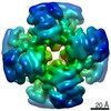 7305MC M: map data used to model this data C: citing same article ( |
|---|---|
| Similar structure data |


-Links
 PDBj
PDBj


- Assembly
| Deposited unit | 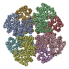
|
|---|---|
| 1 |
|
-Components
| #1: Protein | Mass: 36440.992 Da / Num. of mol.: 8 Source method: isolated from a genetically manipulated source Source: (gene. exp.)  References: UniProt: P62483, Oxidoreductases; Acting on the CH-OH group of donors; With NAD+ or NADP+ as acceptor |
|---|
-Experimental details
-Experiment
| Experiment | Method: ELECTRON MICROSCOPY |
|---|---|
| EM experiment | Aggregation state: PARTICLE / 3D reconstruction method: single particle reconstruction |
- Sample preparation
| Component | Name: Octamer of Kv Beta / Type: COMPLEX / Entity ID: all / Source: RECOMBINANT |
|---|---|
| Source (natural) | Organism: |
| Source (recombinant) | Organism: |
| Buffer solution | pH: 8 |
| Specimen | Embedding applied: NO / Shadowing applied: NO / Staining applied: NO / Vitrification applied: YES |
| Vitrification | Cryogen name: ETHANE |
- Electron microscopy imaging
| Experimental equipment |  Model: Titan Krios / Image courtesy: FEI Company |
|---|---|
| Microscopy | Model: FEI TITAN KRIOS |
| Electron gun | Electron source:  FIELD EMISSION GUN / Accelerating voltage: 300 kV / Illumination mode: FLOOD BEAM FIELD EMISSION GUN / Accelerating voltage: 300 kV / Illumination mode: FLOOD BEAM |
| Electron lens | Mode: BRIGHT FIELD |
| Image recording | Electron dose: 60 e/Å2 / Film or detector model: DIRECT ELECTRON DE-20 (5k x 3k) |
- Processing
| EM software |
| |||||||||
|---|---|---|---|---|---|---|---|---|---|---|
| CTF correction | Type: PHASE FLIPPING AND AMPLITUDE CORRECTION | |||||||||
| Symmetry | Point symmetry: D4 (2x4 fold dihedral) | |||||||||
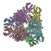
| 3D reconstruction | Resolution: 4.9 Å / Resolution method: FSC 0.143 CUT-OFF / Num. of particles: 24809 / Symmetry type: POINT | |||||||||
| Atomic model building | Protocol: FLEXIBLE FIT / Space: REAL |
