 ムービー
ムービー コントローラー


+ データを開く

- 基本情報
基本情報
| 登録情報 | データベース: PDB / ID: 6c14 | ||||||||||||||||||||||||||||||||||||||||||
|---|---|---|---|---|---|---|---|---|---|---|---|---|---|---|---|---|---|---|---|---|---|---|---|---|---|---|---|---|---|---|---|---|---|---|---|---|---|---|---|---|---|---|---|
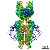
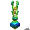
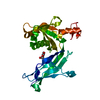
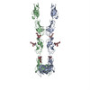
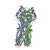
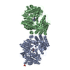
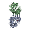
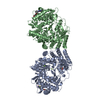
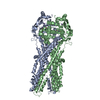
| タイトル | CryoEM structure of mouse PCDH15-1EC-LHFPL5 complex | ||||||||||||||||||||||||||||||||||||||||||
 要素 要素 |
| ||||||||||||||||||||||||||||||||||||||||||
 キーワード キーワード | membrane protein / metal transport / PCDH15 / LHFPL5 / protocadherin / tip link / hair cell / TMHS / hearing | ||||||||||||||||||||||||||||||||||||||||||
| 機能・相同性 |  機能・相同性情報 機能・相同性情報detection of mechanical stimulus involved in equilibrioception / inner ear receptor cell stereocilium organization / stereocilium tip / righting reflex / inner ear auditory receptor cell differentiation / detection of mechanical stimulus involved in sensory perception of sound / stereocilium bundle / stereocilium / non-motile cilium assembly / auditory receptor cell stereocilium organization ...detection of mechanical stimulus involved in equilibrioception / inner ear receptor cell stereocilium organization / stereocilium tip / righting reflex / inner ear auditory receptor cell differentiation / detection of mechanical stimulus involved in sensory perception of sound / stereocilium bundle / stereocilium / non-motile cilium assembly / auditory receptor cell stereocilium organization / adult walking behavior / startle response / homophilic cell adhesion via plasma membrane adhesion molecules / inner ear development / actin filament bundle assembly / photoreceptor outer segment / monoatomic ion transport / visual perception / actin filament organization / morphogenesis of an epithelium / locomotory behavior / sensory perception of sound / multicellular organism growth / response to calcium ion / apical plasma membrane / calcium ion binding / extracellular region / membrane / plasma membrane / cytoplasm 類似検索 - 分子機能 | ||||||||||||||||||||||||||||||||||||||||||
| 生物種 |  | ||||||||||||||||||||||||||||||||||||||||||
| 手法 | 電子顕微鏡法 / 単粒子再構成法 / クライオ電子顕微鏡法 / 解像度: 4.5 Å | ||||||||||||||||||||||||||||||||||||||||||
 データ登録者 データ登録者 | Gouaux, E. / Elferich, J. / Ge, J. | ||||||||||||||||||||||||||||||||||||||||||
 引用 引用 | ジャーナル: Elife / 年: 2018 タイトル: Structure of mouse protocadherin 15 of the stereocilia tip link in complex with LHFPL5. 著者: Jingpeng Ge / Johannes Elferich / April Goehring / Huaying Zhao / Peter Schuck / Eric Gouaux /  要旨: Hearing and balance involve the transduction of mechanical stimuli into electrical signals by deflection of bundles of stereocilia linked together by protocadherin 15 (PCDH15) and cadherin 23 'tip ...Hearing and balance involve the transduction of mechanical stimuli into electrical signals by deflection of bundles of stereocilia linked together by protocadherin 15 (PCDH15) and cadherin 23 'tip links'. PCDH15 transduces tip link tension into opening of a mechano-electrical transduction (MET) ion channel. PCDH15 also interacts with LHFPL5, a candidate subunit of the MET channel. Here we illuminate the PCDH15-LHFPL5 structure, showing how the complex is composed of PCDH15 and LHFPL5 subunit pairs related by a 2-fold axis. The extracellular cadherin domains define a mobile tether coupled to a rigid, 2-fold symmetric 'collar' proximal to the membrane bilayer. LHFPL5 forms extensive interactions with the PCDH15 transmembrane helices and stabilizes the overall PCDH15-LHFPL5 assembly. Our studies illuminate the architecture of the PCDH15-LHFPL5 complex, localize mutations associated with deafness, and shed new light on how forces in the PCDH15 tether may be transduced into the stereocilia membrane. | ||||||||||||||||||||||||||||||||||||||||||
| 履歴 |
|
- 構造の表示
構造の表示
| ムービー |
ムービービューア |
|---|---|
| 構造ビューア | 分子: MolmilJmol/JSmol |
- ダウンロードとリンク
-ダウンロード
| PDBx/mmCIF形式 | 6c14.cif.gz | 202.5 KB | 表示 | PDBx/mmCIF形式 |
|---|---|---|---|---|
| PDB形式 | pdb6c14.ent.gz | 154.1 KB | 表示 | PDB形式 |
| PDBx/mmJSON形式 | 6c14.json.gz | ツリー表示 | PDBx/mmJSON形式 | |
| その他 |  その他のダウンロード その他のダウンロード |
-検証レポート
| 文書・要旨 | 6c14_validation.pdf.gz | 985.4 KB | 表示 | wwPDB検証レポート |
|---|---|---|---|---|
| 文書・詳細版 | 6c14_full_validation.pdf.gz | 990.7 KB | 表示 | |
| XML形式データ | 6c14_validation.xml.gz | 34.2 KB | 表示 | |
| CIF形式データ | 6c14_validation.cif.gz | 51.8 KB | 表示 | |
| アーカイブディレクトリ | https://data.pdbj.org/pub/pdb/validation_reports/c1/6c14ftp://data.pdbj.org/pub/pdb/validation_reports/c1/6c14 | HTTPS FTP |
-関連構造データ


-リンク
 PDBj
PDBj
- 集合体
集合体
| 登録構造単位 | 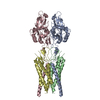
|
|---|---|
| 1 |
|
-要素
| #1: タンパク質 | 分子量: 37458.129 Da / 分子数: 2 / 由来タイプ: 組換発現 / 由来: (組換発現)  Homo sapiens (ヒト) / 参照: UniProt: Q99PJ1 Homo sapiens (ヒト) / 参照: UniProt: Q99PJ1#2: タンパク質 | 分子量: 25067.320 Da / 分子数: 2 / 由来タイプ: 組換発現 / 由来: (組換発現) Homo sapiens (ヒト) / 参照: UniProt: Q4KL25Has protein modification | Y | |
|---|
-実験情報
-実験
| 実験 | 手法: 電子顕微鏡法 |
|---|---|
| EM実験 | 試料の集合状態: PARTICLE / 3次元再構成法: 単粒子再構成法 |
- 試料調製
試料調製
| 構成要素 | 名称: membrane protein complex / タイプ: COMPLEX / Entity ID: all / 由来: RECOMBINANT | ||||||||||||||||||||
|---|---|---|---|---|---|---|---|---|---|---|---|---|---|---|---|---|---|---|---|---|---|
| 由来(天然) | 生物種: | ||||||||||||||||||||
| 由来(組換発現) | 生物種: Homo sapiens (ヒト) | ||||||||||||||||||||
| 緩衝液 | pH: 8 | ||||||||||||||||||||
| 緩衝液成分 |
| ||||||||||||||||||||
| 試料 | 包埋: NO / シャドウイング: NO / 染色: NO / 凍結: YES | ||||||||||||||||||||
| 試料支持 | グリッドの材料: GOLD / グリッドのサイズ: 300 divisions/in. / グリッドのタイプ: Quantifoil R1.2/1.3 | ||||||||||||||||||||
| 急速凍結 | 装置: FEI VITROBOT MARK III / 凍結剤: ETHANE-PROPANE / 湿度: 100 % / 凍結前の試料温度: 285 K |
- 電子顕微鏡撮影
| 実験機器 |  モデル: Titan Krios / 画像提供: FEI Company |
|---|---|
| 顕微鏡 | モデル: FEI TITAN KRIOS |
| 電子銃 | 電子線源:  FIELD EMISSION GUN / 加速電圧: 300 kV / 照射モード: FLOOD BEAM FIELD EMISSION GUN / 加速電圧: 300 kV / 照射モード: FLOOD BEAM |
| 電子レンズ | モード: BRIGHT FIELD |
| 撮影 | 電子線照射量: 74 e/Å2 / 検出モード: SUPER-RESOLUTION フィルム・検出器のモデル: GATAN K2 SUMMIT (4k x 4k) |
- 解析
解析
| ソフトウェア | 名称: PHENIX / バージョン: 1.12_2829: / 分類: 精密化 | ||||||||||||||||||||||||
|---|---|---|---|---|---|---|---|---|---|---|---|---|---|---|---|---|---|---|---|---|---|---|---|---|---|
| EMソフトウェア |
| ||||||||||||||||||||||||
| CTF補正 | タイプ: PHASE FLIPPING AND AMPLITUDE CORRECTION | ||||||||||||||||||||||||
| 対称性 | 点対称性: C2 (2回回転対称) | ||||||||||||||||||||||||
| 3次元再構成 | 解像度: 4.5 Å / 解像度の算出法: FSC 0.143 CUT-OFF / 粒子像の数: 78792 / 対称性のタイプ: POINT | ||||||||||||||||||||||||
| 拘束条件 |
|
