 Movie
Movie Controller


+ Open data

- Basic information
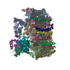
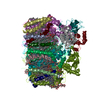
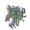
| Entry | Database: PDB / ID: 5y36 | |||||||||
|---|---|---|---|---|---|---|---|---|---|---|
| Title | Cryo-EM structure of SpCas9-sgRNA-DNA ternary complex | |||||||||
 Components Components |
| |||||||||
 Keywords Keywords | HYDROLASE/RNA/DNA / Genome editting / CRIPSR-Cas9 / DNA cleavage mechanism / HYDROLASE-DNA-RNA complex / HYDROLASE-RNA-DNA complex | |||||||||
| Function / homology |  Function and homology information Function and homology informationmaintenance of CRISPR repeat elements / 3'-5' exonuclease activity / DNA endonuclease activity / defense response to virus / Hydrolases; Acting on ester bonds / DNA binding / RNA binding / metal ion binding Similarity search - Function | |||||||||
| Biological species |  Streptococcus pyogenes serotype M1 (bacteria) Streptococcus pyogenes serotype M1 (bacteria) | |||||||||
| Method | ELECTRON MICROSCOPY / single particle reconstruction / cryo EM / Resolution: 5.2 Å | |||||||||
 Authors Authors | Huang, Q. / Li, G. / Huai, C. | |||||||||
| Funding support |  China, 2items China, 2items
| |||||||||
 Citation Citation | Journal: Nat Commun / Year: 2017 Title: Structural insights into DNA cleavage activation of CRISPR-Cas9 system. Authors: Cong Huai / Gan Li / Ruijie Yao / Yingyi Zhang / Mi Cao / Liangliang Kong / Chenqiang Jia / Hui Yuan / Hongyan Chen / Daru Lu / Qiang Huang / Abstract: CRISPR-Cas9 technology has been widely used for genome engineering. Its RNA-guided endonuclease Cas9 binds specifically to target DNA and then cleaves the two DNA strands with HNH and RuvC nuclease ...CRISPR-Cas9 technology has been widely used for genome engineering. Its RNA-guided endonuclease Cas9 binds specifically to target DNA and then cleaves the two DNA strands with HNH and RuvC nuclease domains. However, structural information regarding the DNA cleavage-activating state of two nuclease domains remains sparse. Here, we report a 5.2 Å cryo-EM structure of Cas9 in complex with sgRNA and target DNA. This structure reveals a conformational state of Cas9 in which the HNH domain is closest to the DNA cleavage site. Compared with two known HNH states, our structure shows that the HNH active site moves toward the cleavage site by about 25 and 13 Å, respectively. In combination with EM-based molecular dynamics simulations, we show that residues of the nuclease domains in our structure could form cleavage-compatible conformations with the target DNA. Together, these results strongly suggest that our cryo-EM structure resembles a DNA cleavage-activating architecture of Cas9. | |||||||||
| History |
|
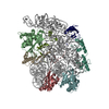
- Structure visualization
| Movie |
Movie viewer |
|---|---|
| Structure viewer | Molecule: MolmilJmol/JSmol |
- Downloads & links
-Download
| PDBx/mmCIF format | 5y36.cif.gz | 340 KB | Display | PDBx/mmCIF format |
|---|---|---|---|---|
| PDB format | pdb5y36.ent.gz | 254.5 KB | Display | PDB format |
| PDBx/mmJSON format | 5y36.json.gz | Tree view | PDBx/mmJSON format | |
| Others |  Other downloads Other downloads |
-Validation report
| Arichive directory | https://data.pdbj.org/pub/pdb/validation_reports/y3/5y36ftp://data.pdbj.org/pub/pdb/validation_reports/y3/5y36 | HTTPS FTP |
|---|
-Related structure data
| Related structure data | 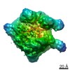 8236MC M: map data used to model this data C: citing same article ( |
|---|---|
| Similar structure data |


-Links
 PDBj
PDBj


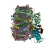
- Assembly
| Deposited unit | 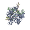
|
|---|---|
| 1 |
|
-Components
| #1: Protein | Mass: 158588.781 Da / Num. of mol.: 1 / Mutation: D10A, H840A Source method: isolated from a genetically manipulated source Source: (gene. exp.) Streptococcus pyogenes serotype M1 (bacteria)Gene: cas9, csn1, SPy_1046 / Plasmid: pET28a Production host: References: UniProt: Q99ZW2, Hydrolases; Acting on ester bonds |
|---|---|
| #2: RNA chain | Mass: 31890.916 Da / Num. of mol.: 1 / Source method: obtained synthetically / Source: (synth.) |
| #3: DNA chain | Mass: 12697.152 Da / Num. of mol.: 1 / Source method: obtained synthetically / Source: (synth.) |
| #4: DNA chain | Mass: 12546.073 Da / Num. of mol.: 1 / Source method: obtained synthetically / Source: (synth.) |
| #5: Chemical |   Mass: 24.305 Da / Num. of mol.: 3 / Source method: obtained synthetically / Formula: Mg Mass: 24.305 Da / Num. of mol.: 3 / Source method: obtained synthetically / Formula: Mg |
-Experimental details
-Experiment
| Experiment | Method: ELECTRON MICROSCOPY |
|---|---|
| EM experiment | Aggregation state: PARTICLE / 3D reconstruction method: single particle reconstruction |
- Sample preparation
| Component |
| |||||||||||||||||||||||||||||||||||
|---|---|---|---|---|---|---|---|---|---|---|---|---|---|---|---|---|---|---|---|---|---|---|---|---|---|---|---|---|---|---|---|---|---|---|---|---|
| Molecular weight |
| |||||||||||||||||||||||||||||||||||
| Source (natural) |
| |||||||||||||||||||||||||||||||||||
| Source (recombinant) |
| |||||||||||||||||||||||||||||||||||
| Buffer solution | pH: 7.5 Details: 20mM Tris-Cl (pH 7.5), 100mM KCl, 5mM MgCl2, 1mM DTT | |||||||||||||||||||||||||||||||||||
| Buffer component |
| |||||||||||||||||||||||||||||||||||
| Specimen | Conc.: 0.45 mg/ml / Embedding applied: NO / Shadowing applied: NO / Staining applied: NO / Vitrification applied: YES Details: dCas9 protein and sgRNA, DNA were cultured at 37 degree centigrade to form a complex, and then monodisperased at 18 degree centigrade overnight. | |||||||||||||||||||||||||||||||||||
| Specimen support | Grid material: COPPER / Grid mesh size: 200 divisions/in. / Grid type: Quantifoil R1.2/1.3 | |||||||||||||||||||||||||||||||||||
| Vitrification | Instrument: FEI VITROBOT MARK IV / Cryogen name: ETHANE / Humidity: 100 % / Chamber temperature: 289 K / Details: blot for 4 seconds before pluging |
- Electron microscopy imaging
| Experimental equipment |  Model: Titan Krios / Image courtesy: FEI Company |
|---|---|
| Microscopy | Model: FEI TITAN KRIOS |
| Electron gun | Electron source:  FIELD EMISSION GUN / Accelerating voltage: 300 kV / Illumination mode: FLOOD BEAM FIELD EMISSION GUN / Accelerating voltage: 300 kV / Illumination mode: FLOOD BEAM |
| Electron lens | Mode: BRIGHT FIELD / Calibrated magnification: 18000 X / Nominal defocus min: 1300 nm / Calibrated defocus max: 3500 nm |
| Specimen holder | Cryogen: NITROGEN / Specimen holder model: FEI TITAN KRIOS AUTOGRID HOLDER |
| Image recording | Electron dose: 10 e/Å2 / Film or detector model: GATAN K2 SUMMIT (4k x 4k) / Num. of real images: 592 |
| Image scans | Width: 3710 / Height: 3838 / Movie frames/image: 38 |
- Processing
| EM software |
| ||||||||||||||||||||||||||||||||||||||||
|---|---|---|---|---|---|---|---|---|---|---|---|---|---|---|---|---|---|---|---|---|---|---|---|---|---|---|---|---|---|---|---|---|---|---|---|---|---|---|---|---|---|
| CTF correction | Type: PHASE FLIPPING ONLY | ||||||||||||||||||||||||||||||||||||||||
| Particle selection | Num. of particles selected: 66845 | ||||||||||||||||||||||||||||||||||||||||
| Symmetry | Point symmetry: C1 (asymmetric) | ||||||||||||||||||||||||||||||||||||||||
| 3D reconstruction | Resolution: 5.2 Å / Resolution method: FSC 0.143 CUT-OFF / Num. of particles: 57484 / Symmetry type: POINT | ||||||||||||||||||||||||||||||||||||||||
| Atomic model building | B value: 80 / Protocol: FLEXIBLE FIT / Space: REAL / Target criteria: Correlation coefficient | ||||||||||||||||||||||||||||||||||||||||
| Atomic model building | PDB-ID: 4OO8 Pdb chain-ID: A / Accession code: 4OO8 / Pdb chain residue range: 1-1368 / Source name: PDB / Type: experimental model |

