 Movie
Movie Controller


+ Open data

- Basic information
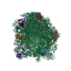
| Entry | Database: PDB / ID: 5xym | ||||||
|---|---|---|---|---|---|---|---|
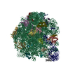
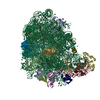
| Title | Large subunit of Mycobacterium smegmatis | ||||||
 Components Components |
| ||||||
 Keywords Keywords | RIBOSOME / Mycobacterium smegmatis ribosome / rRNA / rprotein | ||||||
| Function / homology |  Function and homology information Function and homology informationlarge ribosomal subunit / transferase activity / 5S rRNA binding / ribosomal large subunit assembly / large ribosomal subunit rRNA binding / cytosolic large ribosomal subunit / cytoplasmic translation / tRNA binding / negative regulation of translation / rRNA binding ...large ribosomal subunit / transferase activity / 5S rRNA binding / ribosomal large subunit assembly / large ribosomal subunit rRNA binding / cytosolic large ribosomal subunit / cytoplasmic translation / tRNA binding / negative regulation of translation / rRNA binding / structural constituent of ribosome / ribosome / translation / ribonucleoprotein complex / mRNA binding / cytoplasm Similarity search - Function | ||||||
| Biological species |  Mycobacterium smegmatis (bacteria) Mycobacterium smegmatis (bacteria) | ||||||
| Method | ELECTRON MICROSCOPY / single particle reconstruction / cryo EM / Resolution: 3.08 Å | ||||||
 Authors Authors | Li, Z. / Ge, X. / Zhang, Y. / Zheng, L. / Sanyal, S. / Gao, N. | ||||||
 Citation Citation | Journal: Protein Cell / Year: 2018 Title: Cryo-EM structure of Mycobacterium smegmatis ribosome reveals two unidentified ribosomal proteins close to the functional centers. Authors: Zhifei Li / Xueliang Ge / Yixiao Zhang / Lvqin Zheng / Suparna Sanyal / Ning Gao /   | ||||||
| History |
|
- Structure visualization
| Movie |
Movie viewer |
|---|---|
| Structure viewer | Molecule: MolmilJmol/JSmol |
- Downloads & links
-Download
| PDBx/mmCIF format | 5xym.cif.gz | 2.1 MB | Display | PDBx/mmCIF format |
|---|---|---|---|---|
| PDB format | pdb5xym.ent.gz | 1.6 MB | Display | PDB format |
| PDBx/mmJSON format | 5xym.json.gz | Tree view | PDBx/mmJSON format | |
| Others |  Other downloads Other downloads |
-Validation report
| Arichive directory | https://data.pdbj.org/pub/pdb/validation_reports/xy/5xymftp://data.pdbj.org/pub/pdb/validation_reports/xy/5xym | HTTPS FTP |
|---|
-Related structure data
| Related structure data | 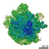 6789MC  6790C 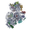 5xyuC M: map data used to model this data C: citing same article ( |
|---|---|
| Similar structure data |


-Links
 PDBj
PDBj


- Assembly
| Deposited unit | 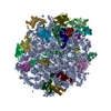
|
|---|---|
| 1 |
|
-Components
+50S ribosomal protein ... , 28 types, 28 molecules 01234CDEFGHJKLMNOPQRSTUVWXYZ
-RNA chain , 2 types, 2 molecules AB
| #6: RNA chain | Mass: 1026283.438 Da / Num. of mol.: 1 / Source method: isolated from a natural source Source: (natural) Mycobacterium smegmatis (strain ATCC 700084 / mc(2)155) (bacteria)Strain: ATCC 700084 / mc(2)155 |
|---|---|
| #7: RNA chain | Mass: 38038.777 Da / Num. of mol.: 1 / Source method: isolated from a natural source Source: (natural) Mycobacterium smegmatis (strain ATCC 700084 / mc(2)155) (bacteria)Strain: ATCC 700084 / mc(2)155 / References: GenBank: 118168627 |
-Protein/peptide / Non-polymers , 2 types, 147 molecules a
| #31: Protein/peptide | Mass: 2841.375 Da / Num. of mol.: 1 / Source method: isolated from a natural source Source: (natural) Mycobacterium smegmatis (strain ATCC 700084 / mc(2)155) (bacteria)Strain: ATCC 700084 / mc(2)155 / References: UniProt: A0QTP4 |
|---|---|
| #32: Chemical | ChemComp-MG /  Mass: 24.305 Da / Num. of mol.: 146 / Source method: obtained synthetically / Formula: Mg Mass: 24.305 Da / Num. of mol.: 146 / Source method: obtained synthetically / Formula: Mg |
-Details
| Has protein modification | Y |
|---|
-Experimental details
-Experiment
| Experiment | Method: ELECTRON MICROSCOPY |
|---|---|
| EM experiment | Aggregation state: PARTICLE / 3D reconstruction method: single particle reconstruction |
- Sample preparation
| Component | Name: Large subunit of Mycobacterium smegmatis ribosome / Type: RIBOSOME / Entity ID: #1-#31 / Source: NATURAL |
|---|---|
| Source (natural) | Organism: Mycobacterium smegmatis (strain ATCC 700084 / mc(2)155) (bacteria) Strain: ATCC 700084 / mc(2)155 |
| Buffer solution | pH: 7.6 |
| Specimen | Embedding applied: NO / Shadowing applied: NO / Staining applied: NO / Vitrification applied: YES |
| Vitrification | Cryogen name: ETHANE |
- Electron microscopy imaging
| Experimental equipment |  Model: Titan Krios / Image courtesy: FEI Company |
|---|---|
| Microscopy | Model: FEI TITAN KRIOS |
| Electron gun | Electron source:  FIELD EMISSION GUN / Accelerating voltage: 300 kV / Illumination mode: FLOOD BEAM FIELD EMISSION GUN / Accelerating voltage: 300 kV / Illumination mode: FLOOD BEAM |
| Electron lens | Mode: BRIGHT FIELD |
| Image recording | Electron dose: 2 e/Å2 / Film or detector model: GATAN K2 SUMMIT (4k x 4k) |
- Processing
| CTF correction | Type: PHASE FLIPPING AND AMPLITUDE CORRECTION |
|---|---|
| 3D reconstruction | Resolution: 3.08 Å / Resolution method: FSC 0.143 CUT-OFF / Num. of particles: 71351 / Symmetry type: POINT |
