 Movie
Movie Controller


+ Open data

- Basic information
| Entry | Database: EMDB / ID: EMD-6790 | |||||||||
|---|---|---|---|---|---|---|---|---|---|---|
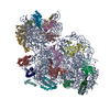
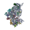
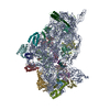
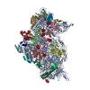
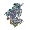
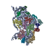
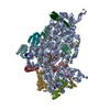
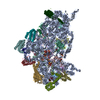
| Title | Small subunit of Mycobacterium smegmatis ribosome | |||||||||
 Map data Map data | ||||||||||
 Sample Sample |
| |||||||||
 Keywords Keywords | Mycobacterium smegmatis ribosome / rRNA / rprotein / RIBOSOME | |||||||||
| Function / homology |  Function and homology information Function and homology informationribosomal small subunit assembly / ribosome biogenesis / ribosomal small subunit biogenesis / small ribosomal subunit / small ribosomal subunit rRNA binding / cytosolic small ribosomal subunit / tRNA binding / rRNA binding / structural constituent of ribosome / ribosome ...ribosomal small subunit assembly / ribosome biogenesis / ribosomal small subunit biogenesis / small ribosomal subunit / small ribosomal subunit rRNA binding / cytosolic small ribosomal subunit / tRNA binding / rRNA binding / structural constituent of ribosome / ribosome / translation / ribonucleoprotein complex / mRNA binding / RNA binding / zinc ion binding / cytoplasm / cytosol Similarity search - Function | |||||||||
| Biological species |  Mycobacterium smegmatis (strain ATCC 700084 / mc(2)155) (bacteria) Mycobacterium smegmatis (strain ATCC 700084 / mc(2)155) (bacteria) | |||||||||
| Method | single particle reconstruction / cryo EM / Resolution: 3.45 Å | |||||||||
 Authors Authors | Li Z / Zhang Y | |||||||||
 Citation Citation | Journal: Protein Cell / Year: 2018 Title: Cryo-EM structure of Mycobacterium smegmatis ribosome reveals two unidentified ribosomal proteins close to the functional centers. Authors: Zhifei Li / Xueliang Ge / Yixiao Zhang / Lvqin Zheng / Suparna Sanyal / Ning Gao /   | |||||||||
| History |
|
- Structure visualization
| Movie |
Movie viewer |
|---|---|
| Structure viewer | EM map: SurfViewMolmilJmol/JSmol |
| Supplemental images |

- Downloads & links
-EMDB archive
| Map data | emd_6790.map.gz | 7.7 MB | EMDB map data format | |
|---|---|---|---|---|
| Header (meta data) | emd-6790-v30.xmlemd-6790.xml | 30.2 KB 30.2 KB | Display Display | EMDB header |
| FSC (resolution estimation) | emd_6790_fsc.xml | 9.8 KB | Display | FSC data file |
| Images | 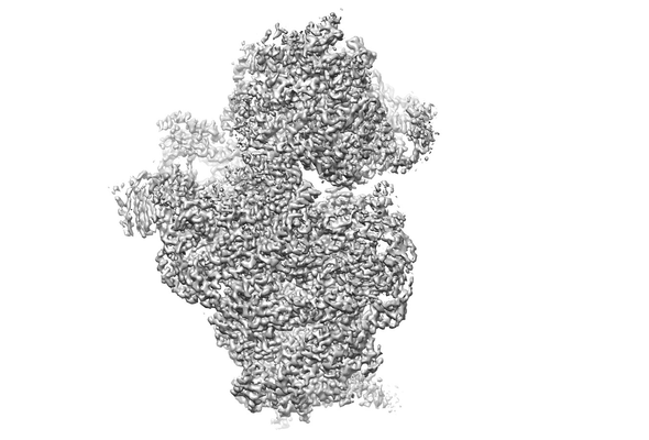 emd_6790.png emd_6790.png | 66.8 KB | ||
| Filedesc metadata | emd-6790.cif.gz | 7.5 KB | ||
| Archive directory |  http://ftp.pdbj.org/pub/emdb/structures/EMD-6790ftp://ftp.pdbj.org/pub/emdb/structures/EMD-6790 http://ftp.pdbj.org/pub/emdb/structures/EMD-6790ftp://ftp.pdbj.org/pub/emdb/structures/EMD-6790 | HTTPS FTP |
-Related structure data
| Related structure data | 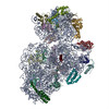 5xyuMC 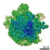 6789C 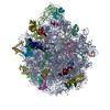 5xymC M: atomic model generated by this map C: citing same article ( |
|---|---|
| Similar structure data |


-Links
| EMDB pages | EMDB (EBI/PDBe) / EMDataResource |
|---|---|
| Related items in Molecule of the Month |


-Map
| File | Download / File: emd_6790.map.gz / Format: CCP4 / Size: 83.7 MB / Type: IMAGE STORED AS FLOATING POINT NUMBER (4 BYTES) | ||||||||||||||||||||||||||||||||||||||||||||||||||||||||||||
|---|---|---|---|---|---|---|---|---|---|---|---|---|---|---|---|---|---|---|---|---|---|---|---|---|---|---|---|---|---|---|---|---|---|---|---|---|---|---|---|---|---|---|---|---|---|---|---|---|---|---|---|---|---|---|---|---|---|---|---|---|---|
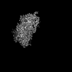
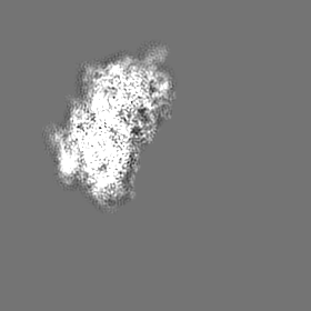
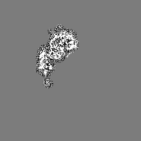
| Projections & slices | Image control
Images are generated by Spider. | ||||||||||||||||||||||||||||||||||||||||||||||||||||||||||||
| Voxel size | X=Y=Z: 1.32 Å | ||||||||||||||||||||||||||||||||||||||||||||||||||||||||||||
| Density |
| ||||||||||||||||||||||||||||||||||||||||||||||||||||||||||||
| Symmetry | Space group: 1 | ||||||||||||||||||||||||||||||||||||||||||||||||||||||||||||
| Details | EMDB XML:
CCP4 map header:
| ||||||||||||||||||||||||||||||||||||||||||||||||||||||||||||
 Z (Sec.)
 Y (Row.)
 X (Col.)


-Supplemental data
- Sample components
+Entire : Small subunit of Mycobacterium smegmatis ribosome
+Supramolecule #1: Small subunit of Mycobacterium smegmatis ribosome
+Macromolecule #1: 16S RNA
+Macromolecule #2: 30S ribosomal protein S3
+Macromolecule #3: 30S ribosomal protein S4
+Macromolecule #4: 30S ribosomal protein S5
+Macromolecule #5: 30S ribosomal protein S6
+Macromolecule #6: 30S ribosomal protein S7
+Macromolecule #7: 30S ribosomal protein S8
+Macromolecule #8: 30S ribosomal protein S9
+Macromolecule #9: 30S ribosomal protein S10
+Macromolecule #10: 30S ribosomal protein S11
+Macromolecule #11: 30S ribosomal protein S12
+Macromolecule #12: 30S ribosomal protein S13
+Macromolecule #13: 30S ribosomal protein S14 type Z
+Macromolecule #14: 30S ribosomal protein S15
+Macromolecule #15: 30S ribosomal protein S16
+Macromolecule #16: 30S ribosomal protein S17
+Macromolecule #17: 30S ribosomal protein S18 2
+Macromolecule #18: 30S ribosomal protein S19
+Macromolecule #19: 30S ribosomal protein S20
+Macromolecule #20: Conserved domain protein
+Macromolecule #21: MAGNESIUM ION
-Experimental details
-Structure determination
| Method | cryo EM |
|---|---|
 Processing Processing | single particle reconstruction |
| Aggregation state | particle |
-Sample preparation
| Buffer | pH: 7.6 |
|---|---|
| Vitrification | Cryogen name: ETHANE |
- Electron microscopy
| Microscope | FEI TITAN KRIOS |
|---|---|
| Image recording | Film or detector model: GATAN K2 SUMMIT (4k x 4k) / Average electron dose: 2.0 e/Å2 |
| Electron beam | Acceleration voltage: 300 kV / Electron source:  FIELD EMISSION GUN FIELD EMISSION GUN |
| Electron optics | Illumination mode: FLOOD BEAM / Imaging mode: BRIGHT FIELD |
| Experimental equipment |  Model: Titan Krios / Image courtesy: FEI Company |

