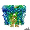
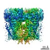
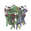
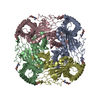
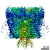
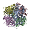
登録情報 データベース : PDB / ID : 5t4dタイトル Cryo-EM structure of Polycystic Kidney Disease protein 2 (PKD2), residues 198-703 hPKD:198-703, Polycystin-2 キーワード / / / / 機能・相同性 分子機能 ドメイン・相同性 構成要素
/ / / / / / / / / / / / / / / / / / / / / / / / / / / / / / / / / / / / / / / / / / / / / / / / / / / / / / / / / / / / / / / / / / / / / / / / / / / / / / / / / / / / / / / / / / / / / / / / / / / / / / / / / / / / / / / / / / / / / 生物種 Homo sapiens (ヒト)手法 / / / 解像度 : 3 Å データ登録者 Shen, P.S. / Yang, X. / DeCaen, P.G. / Liu, X. / Bulkley, D. / Clapham, D.E. / Cao, E. ジャーナル : Cell / 年 : 2016タイトル : The Structure of the Polycystic Kidney Disease Channel PKD2 in Lipid Nanodiscs.著者 : Peter S Shen / Xiaoyong Yang / Paul G DeCaen / Xiaowen Liu / David Bulkley / David E Clapham / Erhu Cao / 要旨 : The Polycystic Kidney Disease 2 (Pkd2) gene is mutated in autosomal dominant polycystic kidney disease (ADPKD), one of the most common human monogenic disorders. Here, we present the cryo-EM ... The Polycystic Kidney Disease 2 (Pkd2) gene is mutated in autosomal dominant polycystic kidney disease (ADPKD), one of the most common human monogenic disorders. Here, we present the cryo-EM structure of PKD2 in lipid bilayers at 3.0 Å resolution, which establishes PKD2 as a homotetrameric ion channel and provides insight into potential mechanisms for its activation. The PKD2 voltage-sensor domain retains two of four gating charges commonly found in those of voltage-gated ion channels. The PKD2 ion permeation pathway is constricted at the selectivity filter and near the cytoplasmic end of S6, suggesting that two gates regulate ion conduction. The extracellular domain of PKD2, a hotspot for ADPKD pathogenic mutations, contributes to channel assembly and strategically interacts with the transmembrane core, likely serving as a physical substrate for extracellular stimuli to allosterically gate the channel. Finally, our structure establishes the molecular basis for the majority of pathogenic mutations in Pkd2-related ADPKD. 履歴 登録 2016年8月29日 登録サイト / 処理サイト 改定 1.0 2016年11月2日 Provider / タイプ 改定 1.1 2016年11月9日 Group 改定 1.2 2016年11月30日 Group 改定 1.3 2017年11月8日 Group / Derived calculations / カテゴリ / pdbx_struct_assemblyItem / _pdbx_struct_assembly.details / _pdbx_struct_assembly.method_details改定 1.4 2019年11月6日 Group / Other / カテゴリ / cellItem _atom_sites.fract_transf_matrix[1][1] / _atom_sites.fract_transf_matrix[2][2] ... _atom_sites.fract_transf_matrix[1][1] / _atom_sites.fract_transf_matrix[2][2] / _atom_sites.fract_transf_matrix[3][3] / _cell.Z_PDB / _cell.length_a / _cell.length_b / _cell.length_c 改定 1.5 2020年7月29日 Group / Derived calculations / Structure summaryカテゴリ chem_comp / entity ... chem_comp / entity / pdbx_chem_comp_identifier / pdbx_entity_nonpoly / struct_conn / struct_site / struct_site_gen Item _chem_comp.name / _chem_comp.type ... _chem_comp.name / _chem_comp.type / _entity.pdbx_description / _pdbx_entity_nonpoly.name / _struct_conn.pdbx_role 解説 / Provider / タイプ
すべて表示 表示を減らす
 ムービー
ムービー コントローラー


 データを開く
 基本情報
基本情報 要素
要素 キーワード
キーワード 機能・相同性情報
 Homo sapiens (ヒト)
 データ登録者
 引用
引用
 構造の表示
構造の表示 ダウンロードとリンク
 その他のダウンロード
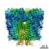


 PDBj
PDBj

 集合体
集合体

 タイプ: D-saccharide, beta linking / 分子量: 221.208 Da / 分子数: 12 / 由来タイプ: 組換発現 / 式: C8H15NO6
 試料調製
試料調製 電子顕微鏡撮影
 FIELD EMISSION GUN / 加速電圧: 300 kV / 照射モード: FLOOD BEAM
 解析
解析