+ データを開く
- 基本情報
基本情報
| 登録情報 | データベース: PDB / ID: 5t0v | ||||||
|---|---|---|---|---|---|---|---|
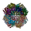
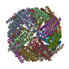
| タイトル | Architecture of the Yeast Mitochondrial Iron-Sulfur Cluster Assembly Machinery: the Sub-Complex Formed by the Iron Donor, Yfh1, and the Scaffold, Isu1 | ||||||
 要素 要素 |
| ||||||
 キーワード キーワード | OXIDOREDUCTASE / Friedreich Ataxia / frataxin / iron-sulfur protein / mitochondria / protein complex | ||||||
| 機能・相同性 |  機能・相同性情報 機能・相同性情報Maturation of TCA enzymes and regulation of TCA cycle / Mitochondrial iron-sulfur cluster biogenesis / Mitochondrial protein import / Complex III assembly / mitochondrial electron transport, succinate to ubiquinone / iron chaperone activity / tRNA wobble uridine modification / iron-sulfur cluster assembly complex / response to iron(II) ion / heme biosynthetic process ...Maturation of TCA enzymes and regulation of TCA cycle / Mitochondrial iron-sulfur cluster biogenesis / Mitochondrial protein import / Complex III assembly / mitochondrial electron transport, succinate to ubiquinone / iron chaperone activity / tRNA wobble uridine modification / iron-sulfur cluster assembly complex / response to iron(II) ion / heme biosynthetic process / iron-sulfur cluster assembly / ferroxidase / ferroxidase activity / ATPase activator activity / glutathione metabolic process / ferric iron binding / ferrous iron binding / mitochondrial intermembrane space / 2 iron, 2 sulfur cluster binding / iron ion transport / intracellular iron ion homeostasis / response to oxidative stress / mitochondrial inner membrane / mitochondrial matrix / iron ion binding / mitochondrion / zinc ion binding / identical protein binding / cytoplasm 類似検索 - 分子機能 | ||||||
| 生物種 |  | ||||||
| 手法 | 電子顕微鏡法 / 単粒子再構成法 / ネガティブ染色法 / 解像度: 17.5 Å | ||||||
 データ登録者 データ登録者 | Ranatunga, W. / Gakh, O. / Galeano, B.K. / Smith IV, D.Y. / Soderberg, C.A. / Al-Karadaghi, S. / Thompson, J.R. / Isaya, G. | ||||||
| 資金援助 |  米国, 1件 米国, 1件
| ||||||
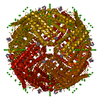
 引用 引用 |  ジャーナル: J Biol Chem / 年: 2016 ジャーナル: J Biol Chem / 年: 2016タイトル: Architecture of the Yeast Mitochondrial Iron-Sulfur Cluster Assembly Machinery: THE SUB-COMPLEX FORMED BY THE IRON DONOR, Yfh1 PROTEIN, AND THE SCAFFOLD, Isu1 PROTEIN. 著者: Wasantha Ranatunga / Oleksandr Gakh / Belinda K Galeano / Douglas Y Smith / Christopher A G Söderberg / Salam Al-Karadaghi / James R Thompson / Grazia Isaya /   要旨: The biosynthesis of Fe-S clusters is a vital process involving the delivery of elemental iron and sulfur to scaffold proteins via molecular interactions that are still poorly defined. We ...The biosynthesis of Fe-S clusters is a vital process involving the delivery of elemental iron and sulfur to scaffold proteins via molecular interactions that are still poorly defined. We reconstituted a stable, functional complex consisting of the iron donor, Yfh1 (yeast frataxin homologue 1), and the Fe-S cluster scaffold, Isu1, with 1:1 stoichiometry, [Yfh1]24·[Isu1]24 Using negative staining transmission EM and single particle analysis, we obtained a three-dimensional reconstruction of this complex at a resolution of ∼17 Å. In addition, via chemical cross-linking, limited proteolysis, and mass spectrometry, we identified protein-protein interaction surfaces within the complex. The data together reveal that [Yfh1]24·[Isu1]24 is a roughly cubic macromolecule consisting of one symmetric Isu1 trimer binding on top of one symmetric Yfh1 trimer at each of its eight vertices. Furthermore, molecular modeling suggests that two subunits of the cysteine desulfurase, Nfs1, may bind symmetrically on top of two adjacent Isu1 trimers in a manner that creates two putative [2Fe-2S] cluster assembly centers. In each center, conserved amino acids known to be involved in sulfur and iron donation by Nfs1 and Yfh1, respectively, are in close proximity to the Fe-S cluster-coordinating residues of Isu1. We suggest that this architecture is suitable to ensure concerted and protected transfer of potentially toxic iron and sulfur atoms to Isu1 during Fe-S cluster assembly. | ||||||
| 履歴 |
|
- 構造の表示
構造の表示
| ムービー |
 ムービービューア ムービービューア |
|---|---|
| 構造ビューア | 分子:  Molmil Molmil Jmol/JSmol Jmol/JSmol |
- ダウンロードとリンク
- ダウンロード
| PDBx/mmCIF形式 |  5t0v.cif.gz 5t0v.cif.gz | 1001.4 KB | 表示 |  PDBx/mmCIF形式 PDBx/mmCIF形式 |
|---|---|---|---|---|
| PDB形式 |  pdb5t0v.ent.gz pdb5t0v.ent.gz | 812.3 KB | 表示 |  PDB形式 PDB形式 |
| PDBx/mmJSON形式 |  5t0v.json.gz 5t0v.json.gz | ツリー表示 |  PDBx/mmJSON形式 PDBx/mmJSON形式 | |
| その他 |  その他のダウンロード その他のダウンロード |
-検証レポート
| 文書・要旨 |  5t0v_validation.pdf.gz 5t0v_validation.pdf.gz | 1.2 MB | 表示 |  wwPDB検証レポート wwPDB検証レポート |
|---|---|---|---|---|
| 文書・詳細版 |  5t0v_full_validation.pdf.gz 5t0v_full_validation.pdf.gz | 1.2 MB | 表示 | |
| XML形式データ |  5t0v_validation.xml.gz 5t0v_validation.xml.gz | 151.4 KB | 表示 | |
| CIF形式データ |  5t0v_validation.cif.gz 5t0v_validation.cif.gz | 238.8 KB | 表示 | |
| アーカイブディレクトリ |  https://data.pdbj.org/pub/pdb/validation_reports/t0/5t0v https://data.pdbj.org/pub/pdb/validation_reports/t0/5t0v ftp://data.pdbj.org/pub/pdb/validation_reports/t0/5t0v ftp://data.pdbj.org/pub/pdb/validation_reports/t0/5t0v | HTTPS FTP |
-関連構造データ
- リンク
リンク
- 集合体
集合体
| 登録構造単位 | 
|
|---|---|
| 1 |
|
- 要素
要素
| #1: タンパク質 | 分子量: 15383.872 Da / 分子数: 24 / 由来タイプ: 組換発現 由来: (組換発現)  遺伝子: ISU1, NUA1, YPL135W / プラスミド: pET28b / 発現宿主:  #2: タンパク質 | 分子量: 13455.976 Da / 分子数: 24 / 由来タイプ: 組換発現 由来: (組換発現)  遺伝子: YFH1, YDL120W / プラスミド: pET24a / 発現宿主:  Has protein modification | Y | |
|---|
-実験情報
-実験
| 実験 | 手法: 電子顕微鏡法 |
|---|---|
| EM実験 | 試料の集合状態: PARTICLE / 3次元再構成法: 単粒子再構成法 |
- 試料調製
試料調製
| 構成要素 | 名称: Yfh1-Isu1 / タイプ: COMPLEX 詳細: macromolecule comprising 24-mer of Yfh1 and 24-mer of Isu1 Entity ID: all / 由来: RECOMBINANT | |||||||||||||||
|---|---|---|---|---|---|---|---|---|---|---|---|---|---|---|---|---|
| 分子量 | 値: 0.7 MDa / 実験値: NO | |||||||||||||||
| 由来(天然) | 生物種:  | |||||||||||||||
| 由来(組換発現) | 生物種:  | |||||||||||||||
| 緩衝液 | pH: 7.3 | |||||||||||||||
| 緩衝液成分 |
| |||||||||||||||
| 試料 | 濃度: 0.12 mg/ml / 包埋: NO / シャドウイング: NO / 染色: YES / 凍結: NO 詳細: The protein complex was prepared by incubating Yfh1 and Isu1 (1:1.5 molar ratio) in HN100 buffer (10 mM HEPES-KOH, pH 7.3, 100 mM NaCl) and purified using Sephacryl S300 gel filtration chromatography. | |||||||||||||||
| 染色 | タイプ: NEGATIVE 詳細: Pre-incubated in HN100 buffer, the grid was placed on an 11-microliter drop of protein sample for 1 minute. Excess protein sample was blotted and washed for 3 seconds by placing the grid on a ...詳細: Pre-incubated in HN100 buffer, the grid was placed on an 11-microliter drop of protein sample for 1 minute. Excess protein sample was blotted and washed for 3 seconds by placing the grid on a drop of sterile water. After excess water was blotted, the grid was stained with 1% w/v uranyl acetate for 1 second and 30 seconds by successively placing it on two separate drops of uranyl acetate, with excess stain drawn off after each step. 染色剤: uranyl acetate | |||||||||||||||
| 試料支持 | 詳細: DV-502A instrument, Denton Vacuum Inc. / グリッドの材料: COPPER / グリッドのサイズ: 400 divisions/in. / グリッドのタイプ: carbon-coated, EMS |
- 電子顕微鏡撮影
| 実験機器 |  モデル: Tecnai F30 / 画像提供: FEI Company |
|---|---|
| 顕微鏡 | モデル: FEI TECNAI F30 |
| 電子銃 | 電子線源:  FIELD EMISSION GUN / 加速電圧: 300 kV / 照射モード: OTHER FIELD EMISSION GUN / 加速電圧: 300 kV / 照射モード: OTHER |
| 電子レンズ | モード: BRIGHT FIELD / 倍率(公称値): 115000 X / 倍率(補正後): 115000 X / 最大 デフォーカス(公称値): 2800 nm / 最小 デフォーカス(公称値): 210 nm / Calibrated defocus min: 210 nm / 最大 デフォーカス(補正後): 2800 nm / Cs: 2 mm / アライメント法: BASIC |
| 試料ホルダ | 凍結剤: NITROGEN / 試料ホルダーモデル: SIDE ENTRY, EUCENTRIC |
| 撮影 | 電子線照射量: 30 e/Å2 フィルム・検出器のモデル: GATAN ULTRASCAN 4000 (4k x 4k) 撮影したグリッド数: 1 / 実像数: 559 |
| 画像スキャン | 横: 4096 / 縦: 4096 |
- 解析
解析
| EMソフトウェア |
| ||||||||||||||||||||||||||||||||||||
|---|---|---|---|---|---|---|---|---|---|---|---|---|---|---|---|---|---|---|---|---|---|---|---|---|---|---|---|---|---|---|---|---|---|---|---|---|---|
| 画像処理 | 詳細: 432 symmetry applied for reconstruction | ||||||||||||||||||||||||||||||||||||
| CTF補正 | 詳細: The ctf.auto function from EMAN2 was applied. / タイプ: PHASE FLIPPING ONLY | ||||||||||||||||||||||||||||||||||||
| 粒子像の選択 | 選択した粒子像数: 4153 | ||||||||||||||||||||||||||||||||||||
| 対称性 | 点対称性: O (正8面体型対称) | ||||||||||||||||||||||||||||||||||||
| 3次元再構成 | 解像度: 17.5 Å / 解像度の算出法: FSC 0.143 CUT-OFF / 粒子像の数: 4153 / アルゴリズム: FOURIER SPACE / 対称性のタイプ: POINT | ||||||||||||||||||||||||||||||||||||
| 原子モデル構築 | プロトコル: RIGID BODY FIT / 空間: REAL |
 ムービー
ムービー コントローラー


 PDBj
PDBj

