 Movie
Movie Controller

[English] 日本語
 Yorodumi
- EMDB-8341: Architecture of the Yeast Mitochondrial Iron-Sulfur Cluster Assem... -
+ Open data

- Basic information
| Entry | Database: EMDB / ID: EMD-8341 | |||||||||
|---|---|---|---|---|---|---|---|---|---|---|
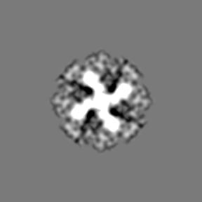
| Title | Architecture of the Yeast Mitochondrial Iron-Sulfur Cluster Assembly Machinery: the Sub-Complex Formed by the Iron Donor, Yfh1, and the Scaffold, Isu1 | |||||||||
 Map data Map data | Yfh1_Isu1 sub-complex | |||||||||
 Sample Sample |
| |||||||||
 Keywords Keywords | Friedreich Ataxia / frataxin / iron-sulfur protein / mitochondria / protein complex / OXIDOREDUCTASE | |||||||||
| Function / homology |  Function and homology information Function and homology informationMitochondrial iron-sulfur cluster biogenesis / Mitochondrial protein import / Maturation of TCA enzymes and regulation of TCA cycle / Complex III assembly / mitochondrial electron transport, succinate to ubiquinone / tRNA wobble uridine modification / iron chaperone activity / iron-sulfur cluster assembly complex / heme biosynthetic process / iron-sulfur cluster assembly ...Mitochondrial iron-sulfur cluster biogenesis / Mitochondrial protein import / Maturation of TCA enzymes and regulation of TCA cycle / Complex III assembly / mitochondrial electron transport, succinate to ubiquinone / tRNA wobble uridine modification / iron chaperone activity / iron-sulfur cluster assembly complex / heme biosynthetic process / iron-sulfur cluster assembly / response to iron(II) ion / ferroxidase / ATPase activator activity / ferroxidase activity / ferric iron binding / glutathione metabolic process / iron ion transport / ferrous iron binding / mitochondrial intermembrane space / 2 iron, 2 sulfur cluster binding / response to oxidative stress / intracellular iron ion homeostasis / mitochondrial inner membrane / iron ion binding / mitochondrial matrix / mitochondrion / zinc ion binding / identical protein binding / cytoplasm Similarity search - Function | |||||||||
| Biological species |  | |||||||||
| Method | single particle reconstruction / negative staining / Resolution: 17.5 Å | |||||||||
 Authors Authors | Ranatunga W / Gakh O | |||||||||
| Funding support |  United States, 1 items United States, 1 items
| |||||||||
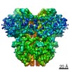
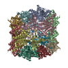
 Citation Citation | Journal: J Biol Chem / Year: 2016 Title: Architecture of the Yeast Mitochondrial Iron-Sulfur Cluster Assembly Machinery: THE SUB-COMPLEX FORMED BY THE IRON DONOR, Yfh1 PROTEIN, AND THE SCAFFOLD, Isu1 PROTEIN. Authors: Wasantha Ranatunga / Oleksandr Gakh / Belinda K Galeano / Douglas Y Smith / Christopher A G Söderberg / Salam Al-Karadaghi / James R Thompson / Grazia Isaya /  Abstract: The biosynthesis of Fe-S clusters is a vital process involving the delivery of elemental iron and sulfur to scaffold proteins via molecular interactions that are still poorly defined. We ...The biosynthesis of Fe-S clusters is a vital process involving the delivery of elemental iron and sulfur to scaffold proteins via molecular interactions that are still poorly defined. We reconstituted a stable, functional complex consisting of the iron donor, Yfh1 (yeast frataxin homologue 1), and the Fe-S cluster scaffold, Isu1, with 1:1 stoichiometry, [Yfh1]24·[Isu1]24 Using negative staining transmission EM and single particle analysis, we obtained a three-dimensional reconstruction of this complex at a resolution of ∼17 Å. In addition, via chemical cross-linking, limited proteolysis, and mass spectrometry, we identified protein-protein interaction surfaces within the complex. The data together reveal that [Yfh1]24·[Isu1]24 is a roughly cubic macromolecule consisting of one symmetric Isu1 trimer binding on top of one symmetric Yfh1 trimer at each of its eight vertices. Furthermore, molecular modeling suggests that two subunits of the cysteine desulfurase, Nfs1, may bind symmetrically on top of two adjacent Isu1 trimers in a manner that creates two putative [2Fe-2S] cluster assembly centers. In each center, conserved amino acids known to be involved in sulfur and iron donation by Nfs1 and Yfh1, respectively, are in close proximity to the Fe-S cluster-coordinating residues of Isu1. We suggest that this architecture is suitable to ensure concerted and protected transfer of potentially toxic iron and sulfur atoms to Isu1 during Fe-S cluster assembly. | |||||||||
| History |
|
- Structure visualization
| Movie |
Movie viewer |
|---|---|
| Structure viewer | EM map: SurfViewMolmilJmol/JSmol |
| Supplemental images |

- Downloads & links
-EMDB archive
| Map data | emd_8341.map.gz | 11 MB | EMDB map data format | |
|---|---|---|---|---|
| Header (meta data) | emd-8341-v30.xmlemd-8341.xml | 14.8 KB 14.8 KB | Display Display | EMDB header |
| FSC (resolution estimation) | emd_8341_fsc.xml | 12 KB | Display | FSC data file |
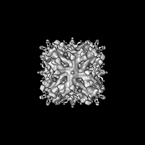
| Images | 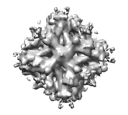 emd_8341.png emd_8341.png | 67.1 KB | ||
| Filedesc metadata | emd-8341.cif.gz | 6.1 KB | ||
| Archive directory |  http://ftp.pdbj.org/pub/emdb/structures/EMD-8341ftp://ftp.pdbj.org/pub/emdb/structures/EMD-8341 http://ftp.pdbj.org/pub/emdb/structures/EMD-8341ftp://ftp.pdbj.org/pub/emdb/structures/EMD-8341 | HTTPS FTP |
-Related structure data
| Related structure data |  5t0vMC M: atomic model generated by this map C: citing same article ( |
|---|---|
| Similar structure data |

-Links
| EMDB pages | EMDB (EBI/PDBe) / EMDataResource |
|---|---|
| Related items in Molecule of the Month |


-Map
| File | Download / File: emd_8341.map.gz / Format: CCP4 / Size: 91.1 MB / Type: IMAGE STORED AS FLOATING POINT NUMBER (4 BYTES) | ||||||||||||||||||||||||||||||||||||||||||||||||||||||||||||||||||||
|---|---|---|---|---|---|---|---|---|---|---|---|---|---|---|---|---|---|---|---|---|---|---|---|---|---|---|---|---|---|---|---|---|---|---|---|---|---|---|---|---|---|---|---|---|---|---|---|---|---|---|---|---|---|---|---|---|---|---|---|---|---|---|---|---|---|---|---|---|---|
| Annotation | Yfh1_Isu1 sub-complex | ||||||||||||||||||||||||||||||||||||||||||||||||||||||||||||||||||||
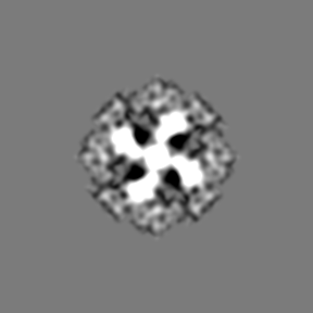
| Projections & slices | Image control
Images are generated by Spider. | ||||||||||||||||||||||||||||||||||||||||||||||||||||||||||||||||||||
| Voxel size | X=Y=Z: 1.034 Å | ||||||||||||||||||||||||||||||||||||||||||||||||||||||||||||||||||||
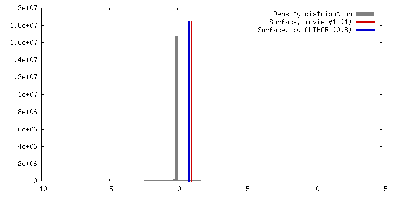
| Density |
| ||||||||||||||||||||||||||||||||||||||||||||||||||||||||||||||||||||
| Symmetry | Space group: 1 | ||||||||||||||||||||||||||||||||||||||||||||||||||||||||||||||||||||
| Details | EMDB XML:
CCP4 map header:
| ||||||||||||||||||||||||||||||||||||||||||||||||||||||||||||||||||||
 Z (Sec.)
 Y (Row.)
 X (Col.)


-Supplemental data
- Sample components
-Entire : Yfh1-Isu1
| Entire | Name: Yfh1-Isu1 |
|---|---|
| Components |
|
-Supramolecule #1: Yfh1-Isu1
| Supramolecule | Name: Yfh1-Isu1 / type: complex / ID: 1 / Parent: 0 / Macromolecule list: all Details: macromolecule comprising 24-mer of Yfh1 and 24-mer of Isu1 |
|---|---|
| Source (natural) | Organism: |
| Molecular weight | Theoretical: 700 KDa |
-Macromolecule #1: Iron sulfur cluster assembly protein 1, mitochondrial
| Macromolecule | Name: Iron sulfur cluster assembly protein 1, mitochondrial / type: protein_or_peptide / ID: 1 / Number of copies: 24 / Enantiomer: LEVO |
|---|---|
| Source (natural) | Organism: |
| Molecular weight | Theoretical: 15.383872 KDa |
| Recombinant expression | Organism:  |
| Sequence | String: GSHMSSITKR LYHPKVIEHY THPRNVGSLD KKLPNVGTGL VGAPACGDVM RLQIKVNDST GVIEDVKFKT FGCGSAIASS SYMTELVQG MTLDDAAKIK NTEIAKELSL PPVKLHCSML AEDAIKAAIK DYKSKRNTPT MLS UniProtKB: Iron sulfur cluster assembly protein 1, mitochondrial |
-Macromolecule #2: Frataxin homolog, mitochondrial
| Macromolecule | Name: Frataxin homolog, mitochondrial / type: protein_or_peptide / ID: 2 / Number of copies: 24 / Enantiomer: LEVO / EC number: ferroxidase |
|---|---|
| Source (natural) | Organism: |
| Molecular weight | Theoretical: 13.455976 KDa |
| Recombinant expression | Organism: |
| Sequence | String: VESSTDGQVV PQEVLNLPLE KAHEEADDYL DHLLDSLEEL SEAHPDCIPD VELSHGVMTL EIPAFGTYVI NKQPPNKQIW LASPLSGPN RFDLLNGEWV SLRNGTKLTD ILTEEVEKAI SK UniProtKB: Frataxin homolog, mitochondrial |
-Experimental details
-Structure determination
| Method | negative staining |
|---|---|
 Processing Processing | single particle reconstruction |
| Aggregation state | particle |
-Sample preparation
| Concentration | 0.12 mg/mL | |||||||||
|---|---|---|---|---|---|---|---|---|---|---|
| Buffer | pH: 7.3 Component:
| |||||||||
| Staining | Type: NEGATIVE / Material: uranyl acetate Details: Pre-incubated in HN100 buffer, the grid was placed on an 11-microliter drop of protein sample for 1 minute. Excess protein sample was blotted and washed for 3 seconds by placing the grid on ...Details: Pre-incubated in HN100 buffer, the grid was placed on an 11-microliter drop of protein sample for 1 minute. Excess protein sample was blotted and washed for 3 seconds by placing the grid on a drop of sterile water. After excess water was blotted, the grid was stained with 1% w/v uranyl acetate for 1 second and 30 seconds by successively placing it on two separate drops of uranyl acetate, with excess stain drawn off after each step. | |||||||||
| Grid | Model: carbon-coated, EMS / Material: COPPER / Mesh: 400 / Pretreatment - Type: GLOW DISCHARGE / Pretreatment - Time: 30 sec. / Details: DV-502A instrument, Denton Vacuum Inc. | |||||||||
| Details | The protein complex was prepared by incubating Yfh1 and Isu1 (1:1.5 molar ratio) in HN100 buffer (10 mM HEPES-KOH, pH 7.3, 100 mM NaCl) and purified using Sephacryl S300 gel filtration chromatography. |
- Electron microscopy
| Microscope | FEI TECNAI F30 |
|---|---|
| Image recording | Film or detector model: GATAN ULTRASCAN 4000 (4k x 4k) / Digitization - Dimensions - Width: 4096 pixel / Digitization - Dimensions - Height: 4096 pixel / Number grids imaged: 1 / Number real images: 559 / Average electron dose: 30.0 e/Å2 |
| Electron beam | Acceleration voltage: 300 kV / Electron source:  FIELD EMISSION GUN FIELD EMISSION GUN |
| Electron optics | Calibrated defocus max: 2.8000000000000003 µm / Calibrated defocus min: 0.21 µm / Calibrated magnification: 115000 / Illumination mode: OTHER / Imaging mode: BRIGHT FIELD / Cs: 2.0 mm / Nominal defocus max: 2.8000000000000003 µm / Nominal defocus min: 0.21 µm / Nominal magnification: 115000 |
| Sample stage | Specimen holder model: SIDE ENTRY, EUCENTRIC / Cooling holder cryogen: NITROGEN |
| Experimental equipment |  Model: Tecnai F30 / Image courtesy: FEI Company |
+Image processing

-Atomic model buiding 1
| Refinement | Space: REAL / Protocol: RIGID BODY FIT |
|---|---|
| Output model | PDB-5t0v: |
