 Movie
Movie Controller

[English] 日本語
 Yorodumi
- PDB-5t0v: Architecture of the Yeast Mitochondrial Iron-Sulfur Cluster Assem... -
+ Open data

- Basic information
| Entry | Database: PDB / ID: 5t0v | ||||||
|---|---|---|---|---|---|---|---|
| Title | Architecture of the Yeast Mitochondrial Iron-Sulfur Cluster Assembly Machinery: the Sub-Complex Formed by the Iron Donor, Yfh1, and the Scaffold, Isu1 | ||||||
 Components Components |
| ||||||
 Keywords Keywords | OXIDOREDUCTASE / Friedreich Ataxia / frataxin / iron-sulfur protein / mitochondria / protein complex | ||||||
| Function / homology |  Function and homology information Function and homology informationMitochondrial iron-sulfur cluster biogenesis / Mitochondrial protein import / Maturation of TCA enzymes and regulation of TCA cycle / Complex III assembly / mitochondrial electron transport, succinate to ubiquinone / tRNA wobble uridine modification / iron chaperone activity / iron-sulfur cluster assembly complex / heme biosynthetic process / iron-sulfur cluster assembly ...Mitochondrial iron-sulfur cluster biogenesis / Mitochondrial protein import / Maturation of TCA enzymes and regulation of TCA cycle / Complex III assembly / mitochondrial electron transport, succinate to ubiquinone / tRNA wobble uridine modification / iron chaperone activity / iron-sulfur cluster assembly complex / heme biosynthetic process / iron-sulfur cluster assembly / response to iron(II) ion / ferroxidase / ATPase activator activity / ferroxidase activity / ferric iron binding / glutathione metabolic process / iron ion transport / ferrous iron binding / mitochondrial intermembrane space / 2 iron, 2 sulfur cluster binding / response to oxidative stress / intracellular iron ion homeostasis / mitochondrial inner membrane / iron ion binding / mitochondrial matrix / mitochondrion / zinc ion binding / identical protein binding / cytoplasm Similarity search - Function | ||||||
| Biological species |  | ||||||
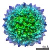
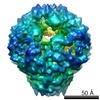
| Method | ELECTRON MICROSCOPY / single particle reconstruction / negative staining / Resolution: 17.5 Å | ||||||
 Authors Authors | Ranatunga, W. / Gakh, O. / Galeano, B.K. / Smith IV, D.Y. / Soderberg, C.A. / Al-Karadaghi, S. / Thompson, J.R. / Isaya, G. | ||||||
| Funding support |  United States, 1items United States, 1items
| ||||||
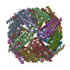
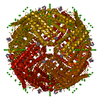
 Citation Citation | Journal: J Biol Chem / Year: 2016 Title: Architecture of the Yeast Mitochondrial Iron-Sulfur Cluster Assembly Machinery: THE SUB-COMPLEX FORMED BY THE IRON DONOR, Yfh1 PROTEIN, AND THE SCAFFOLD, Isu1 PROTEIN. Authors: Wasantha Ranatunga / Oleksandr Gakh / Belinda K Galeano / Douglas Y Smith / Christopher A G Söderberg / Salam Al-Karadaghi / James R Thompson / Grazia Isaya /  Abstract: The biosynthesis of Fe-S clusters is a vital process involving the delivery of elemental iron and sulfur to scaffold proteins via molecular interactions that are still poorly defined. We ...The biosynthesis of Fe-S clusters is a vital process involving the delivery of elemental iron and sulfur to scaffold proteins via molecular interactions that are still poorly defined. We reconstituted a stable, functional complex consisting of the iron donor, Yfh1 (yeast frataxin homologue 1), and the Fe-S cluster scaffold, Isu1, with 1:1 stoichiometry, [Yfh1]24·[Isu1]24 Using negative staining transmission EM and single particle analysis, we obtained a three-dimensional reconstruction of this complex at a resolution of ∼17 Å. In addition, via chemical cross-linking, limited proteolysis, and mass spectrometry, we identified protein-protein interaction surfaces within the complex. The data together reveal that [Yfh1]24·[Isu1]24 is a roughly cubic macromolecule consisting of one symmetric Isu1 trimer binding on top of one symmetric Yfh1 trimer at each of its eight vertices. Furthermore, molecular modeling suggests that two subunits of the cysteine desulfurase, Nfs1, may bind symmetrically on top of two adjacent Isu1 trimers in a manner that creates two putative [2Fe-2S] cluster assembly centers. In each center, conserved amino acids known to be involved in sulfur and iron donation by Nfs1 and Yfh1, respectively, are in close proximity to the Fe-S cluster-coordinating residues of Isu1. We suggest that this architecture is suitable to ensure concerted and protected transfer of potentially toxic iron and sulfur atoms to Isu1 during Fe-S cluster assembly. | ||||||
| History |
|
- Structure visualization
| Movie |
Movie viewer |
|---|---|
| Structure viewer | Molecule: MolmilJmol/JSmol |
- Downloads & links
-Download
| PDBx/mmCIF format | 5t0v.cif.gz | 1001.4 KB | Display | PDBx/mmCIF format |
|---|---|---|---|---|
| PDB format | pdb5t0v.ent.gz | 812.3 KB | Display | PDB format |
| PDBx/mmJSON format | 5t0v.json.gz | Tree view | PDBx/mmJSON format | |
| Others |  Other downloads Other downloads |
-Validation report
| Arichive directory | https://data.pdbj.org/pub/pdb/validation_reports/t0/5t0vftp://data.pdbj.org/pub/pdb/validation_reports/t0/5t0v | HTTPS FTP |
|---|
-Related structure data
| Related structure data |  8341MC M: map data used to model this data C: citing same article ( |
|---|---|
| Similar structure data |


-Links
 PDBj
PDBj

- Assembly
| Deposited unit | 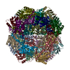
|
|---|---|
| 1 |
|
-Components
| #1: Protein | Mass: 15383.872 Da / Num. of mol.: 24 Source method: isolated from a genetically manipulated source Source: (gene. exp.) Gene: ISU1, NUA1, YPL135W / Plasmid: pET28b / Production host:  #2: Protein | Mass: 13455.976 Da / Num. of mol.: 24 Source method: isolated from a genetically manipulated source Source: (gene. exp.) Gene: YFH1, YDL120W / Plasmid: pET24a / Production host: Has protein modification | Y | |
|---|
-Experimental details
-Experiment
| Experiment | Method: ELECTRON MICROSCOPY |
|---|---|
| EM experiment | Aggregation state: PARTICLE / 3D reconstruction method: single particle reconstruction |
- Sample preparation
| Component | Name: Yfh1-Isu1 / Type: COMPLEX Details: macromolecule comprising 24-mer of Yfh1 and 24-mer of Isu1 Entity ID: all / Source: RECOMBINANT | |||||||||||||||
|---|---|---|---|---|---|---|---|---|---|---|---|---|---|---|---|---|
| Molecular weight | Value: 0.7 MDa / Experimental value: NO | |||||||||||||||
| Source (natural) | Organism: | |||||||||||||||
| Source (recombinant) | Organism: | |||||||||||||||
| Buffer solution | pH: 7.3 | |||||||||||||||
| Buffer component |
| |||||||||||||||
| Specimen | Conc.: 0.12 mg/ml / Embedding applied: NO / Shadowing applied: NO / Staining applied: YES / Vitrification applied: NO Details: The protein complex was prepared by incubating Yfh1 and Isu1 (1:1.5 molar ratio) in HN100 buffer (10 mM HEPES-KOH, pH 7.3, 100 mM NaCl) and purified using Sephacryl S300 gel filtration chromatography. | |||||||||||||||
| EM staining | Type: NEGATIVE Details: Pre-incubated in HN100 buffer, the grid was placed on an 11-microliter drop of protein sample for 1 minute. Excess protein sample was blotted and washed for 3 seconds by placing the grid on ...Details: Pre-incubated in HN100 buffer, the grid was placed on an 11-microliter drop of protein sample for 1 minute. Excess protein sample was blotted and washed for 3 seconds by placing the grid on a drop of sterile water. After excess water was blotted, the grid was stained with 1% w/v uranyl acetate for 1 second and 30 seconds by successively placing it on two separate drops of uranyl acetate, with excess stain drawn off after each step. Material: uranyl acetate | |||||||||||||||
| Specimen support | Details: DV-502A instrument, Denton Vacuum Inc. / Grid material: COPPER / Grid mesh size: 400 divisions/in. / Grid type: carbon-coated, EMS |
- Electron microscopy imaging
| Experimental equipment |  Model: Tecnai F30 / Image courtesy: FEI Company |
|---|---|
| Microscopy | Model: FEI TECNAI F30 |
| Electron gun | Electron source:  FIELD EMISSION GUN / Accelerating voltage: 300 kV / Illumination mode: OTHER FIELD EMISSION GUN / Accelerating voltage: 300 kV / Illumination mode: OTHER |
| Electron lens | Mode: BRIGHT FIELD / Nominal magnification: 115000 X / Calibrated magnification: 115000 X / Nominal defocus max: 2800 nm / Nominal defocus min: 210 nm / Calibrated defocus min: 210 nm / Calibrated defocus max: 2800 nm / Cs: 2 mm / Alignment procedure: BASIC |
| Specimen holder | Cryogen: NITROGEN / Specimen holder model: SIDE ENTRY, EUCENTRIC |
| Image recording | Electron dose: 30 e/Å2 / Film or detector model: GATAN ULTRASCAN 4000 (4k x 4k) / Num. of grids imaged: 1 / Num. of real images: 559 |
| Image scans | Width: 4096 / Height: 4096 |
- Processing
| EM software |
| ||||||||||||||||||||||||||||||||||||
|---|---|---|---|---|---|---|---|---|---|---|---|---|---|---|---|---|---|---|---|---|---|---|---|---|---|---|---|---|---|---|---|---|---|---|---|---|---|
| Image processing | Details: 432 symmetry applied for reconstruction | ||||||||||||||||||||||||||||||||||||
| CTF correction | Details: The ctf.auto function from EMAN2 was applied. / Type: PHASE FLIPPING ONLY | ||||||||||||||||||||||||||||||||||||
| Particle selection | Num. of particles selected: 4153 | ||||||||||||||||||||||||||||||||||||
| Symmetry | Point symmetry: O (octahedral) | ||||||||||||||||||||||||||||||||||||
| 3D reconstruction | Resolution: 17.5 Å / Resolution method: FSC 0.143 CUT-OFF / Num. of particles: 4153 / Algorithm: FOURIER SPACE / Symmetry type: POINT | ||||||||||||||||||||||||||||||||||||
| Atomic model building | Protocol: RIGID BODY FIT / Space: REAL |
