 Movie
Movie Controller

[English] 日本語
 Yorodumi
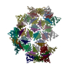
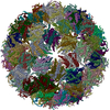
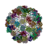
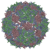
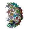
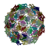
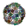
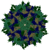
- PDB-5lqp: Cryo-EM reconstruction of bacteriophage AP205 virus-like particles. -
+ Open data

- Basic information
| Entry | Database: PDB / ID: 5lqp | ||||||
|---|---|---|---|---|---|---|---|
| Title | Cryo-EM reconstruction of bacteriophage AP205 virus-like particles. | ||||||
 Components Components | Coat protein | ||||||
 Keywords Keywords | VIRUS LIKE PARTICLE / RNA bacteriophage / Leviviridae / coat protein / virus-like particle | ||||||
| Function / homology | : / AP205 coat protein / viral capsid / Coat protein Function and homology information Function and homology information | ||||||
| Biological species |  Acinetobacter phage AP205 (virus) Acinetobacter phage AP205 (virus) | ||||||
| Method | ELECTRON MICROSCOPY / single particle reconstruction / cryo EM / Resolution: 6 Å | ||||||
 Authors Authors | Diebolder, C.A. / Rumnieks, J. / Tars, K. / Koning, R.I. | ||||||
 Citation Citation | Journal: J Mol Biol / Year: 2016 Title: Structure of AP205 Coat Protein Reveals Circular Permutation in ssRNA Bacteriophages. Authors: Mihails Shishovs / Janis Rumnieks / Christoph Diebolder / Kristaps Jaudzems / Loren B Andreas / Jan Stanek / Andris Kazaks / Svetlana Kotelovica / Inara Akopjana / Guido Pintacuda / Roman I ...Authors: Mihails Shishovs / Janis Rumnieks / Christoph Diebolder / Kristaps Jaudzems / Loren B Andreas / Jan Stanek / Andris Kazaks / Svetlana Kotelovica / Inara Akopjana / Guido Pintacuda / Roman I Koning / Kaspars Tars /    Abstract: AP205 is a single-stranded RNA bacteriophage that has a coat protein sequence not similar to any other known single-stranded RNA phage. Here, we report an atomic-resolution model of the AP205 virus- ...AP205 is a single-stranded RNA bacteriophage that has a coat protein sequence not similar to any other known single-stranded RNA phage. Here, we report an atomic-resolution model of the AP205 virus-like particle based on a crystal structure of an unassembled coat protein dimer and a cryo-electron microscopy reconstruction of the assembled particle, together with secondary structure information from site-specific solid-state NMR data. The AP205 coat protein dimer adopts the conserved Leviviridae coat protein fold except for the N-terminal region, which forms a beta-hairpin in the other known single-stranded RNA phages. AP205 has a similar structure at the same location formed by N- and C-terminal beta-strands, making it a circular permutant compared to the other coat proteins. The permutation moves the coat protein termini to the most surface-exposed part of the assembled particle, which explains its increased tolerance to long N- and C-terminal fusions. | ||||||
| History |
|
- Structure visualization
| Movie |
Movie viewer |
|---|---|
| Structure viewer | Molecule: MolmilJmol/JSmol |
- Downloads & links
-Download
| PDBx/mmCIF format | 5lqp.cif.gz | 3.5 MB | Display | PDBx/mmCIF format |
|---|---|---|---|---|
| PDB format | pdb5lqp.ent.gz | Display | PDB format | |
| PDBx/mmJSON format | 5lqp.json.gz | Tree view | PDBx/mmJSON format | |
| Others |  Other downloads Other downloads |
-Validation report
| Arichive directory | https://data.pdbj.org/pub/pdb/validation_reports/lq/5lqpftp://data.pdbj.org/pub/pdb/validation_reports/lq/5lqp | HTTPS FTP |
|---|
-Related structure data
| Related structure data |  4098MC 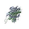 5fs4C M: map data used to model this data C: citing same article ( |
|---|---|
| Similar structure data |
-Links
 PDBj
PDBj- Assembly
| Deposited unit | 
|
|---|---|
| 1 |
|
-Components
| #1: Protein | Mass: 13820.569 Da / Num. of mol.: 180 Source method: isolated from a genetically manipulated source Source: (gene. exp.) Acinetobacter phage AP205 (virus) / Production host:  Has protein modification | Y | |
|---|
-Experimental details
-Experiment
| Experiment | Method: ELECTRON MICROSCOPY |
|---|---|
| EM experiment | Aggregation state: PARTICLE / 3D reconstruction method: single particle reconstruction |
- Sample preparation
| Component | Name: AP205 / Type: VIRUS / Entity ID: all / Source: NATURAL |
|---|---|
| Source (natural) | Organism: Acinetobacter phage AP205 (virus) |
| Details of virus | Empty: NO / Enveloped: NO / Isolate: SPECIES / Type: VIRUS-LIKE PARTICLE |
| Buffer solution | pH: 8 |
| Specimen | Embedding applied: NO / Shadowing applied: NO / Staining applied: NO / Vitrification applied: YES |
| Specimen support | Grid material: COPPER / Grid mesh size: 400 divisions/in. / Grid type: Quantifoil R2/1 |
| Vitrification | Instrument: FEI VITROBOT MARK IV / Cryogen name: ETHANE / Humidity: 95 % / Chamber temperature: 298 K |
- Electron microscopy imaging
| Experimental equipment |  Model: Titan Krios / Image courtesy: FEI Company |
|---|---|
| Microscopy | Model: FEI TITAN KRIOS |
| Electron gun | Electron source:  FIELD EMISSION GUN / Accelerating voltage: 300 kV / Illumination mode: FLOOD BEAM FIELD EMISSION GUN / Accelerating voltage: 300 kV / Illumination mode: FLOOD BEAM |
| Electron lens | Mode: BRIGHT FIELD / Nominal magnification: 59000 X / Nominal defocus max: 2000 nm / Nominal defocus min: 1000 nm / Calibrated defocus min: 1000 nm / Calibrated defocus max: 2500 nm / Cs: 2.7 mm / Alignment procedure: COMA FREE |
| Specimen holder | Cryogen: NITROGEN / Specimen holder model: FEI TITAN KRIOS AUTOGRID HOLDER |
| Image recording | Electron dose: 8 e/Å2 / Detector mode: INTEGRATING / Film or detector model: FEI FALCON II (4k x 4k) |
| EM imaging optics | Energyfilter name: FEI SFEG |
| Image scans | Movie frames/image: 7 / Used frames/image: 1-7 |
- Processing
| EM software |
| ||||||||||||||||||||||||||||||||
|---|---|---|---|---|---|---|---|---|---|---|---|---|---|---|---|---|---|---|---|---|---|---|---|---|---|---|---|---|---|---|---|---|---|
| CTF correction | Type: PHASE FLIPPING AND AMPLITUDE CORRECTION | ||||||||||||||||||||||||||||||||
| Particle selection | Num. of particles selected: 63253 | ||||||||||||||||||||||||||||||||
| Symmetry | Point symmetry: I (icosahedral) | ||||||||||||||||||||||||||||||||
| 3D reconstruction | Resolution: 6 Å / Resolution method: FSC 0.143 CUT-OFF / Num. of particles: 2215 / Symmetry type: POINT |
