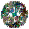
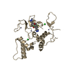
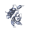
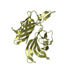
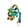
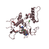
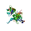
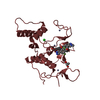
Journal: J Mol Biol / Year: 2016 Title: Structure of AP205 Coat Protein Reveals Circular Permutation in ssRNA Bacteriophages. Authors: Mihails Shishovs / Janis Rumnieks / Christoph Diebolder / Kristaps Jaudzems / Loren B Andreas / Jan Stanek / Andris Kazaks / Svetlana Kotelovica / Inara Akopjana / Guido Pintacuda / Roman I ...Authors: Mihails Shishovs / Janis Rumnieks / Christoph Diebolder / Kristaps Jaudzems / Loren B Andreas / Jan Stanek / Andris Kazaks / Svetlana Kotelovica / Inara Akopjana / Guido Pintacuda / Roman I Koning / Kaspars Tars / Abstract: AP205 is a single-stranded RNA bacteriophage that has a coat protein sequence not similar to any other known single-stranded RNA phage. Here, we report an atomic-resolution model of the AP205 virus- ...AP205 is a single-stranded RNA bacteriophage that has a coat protein sequence not similar to any other known single-stranded RNA phage. Here, we report an atomic-resolution model of the AP205 virus-like particle based on a crystal structure of an unassembled coat protein dimer and a cryo-electron microscopy reconstruction of the assembled particle, together with secondary structure information from site-specific solid-state NMR data. The AP205 coat protein dimer adopts the conserved Leviviridae coat protein fold except for the N-terminal region, which forms a beta-hairpin in the other known single-stranded RNA phages. AP205 has a similar structure at the same location formed by N- and C-terminal beta-strands, making it a circular permutant compared to the other coat proteins. The permutation moves the coat protein termini to the most surface-exposed part of the assembled particle, which explains its increased tolerance to long N- and C-terminal fusions.
History
Deposition
Dec 29, 2015
Deposition site: PDBE / Processing site: PDBE
Revision 1.0
Sep 21, 2016
Provider: repository / Type: Initial release
Revision 1.1
Nov 9, 2016
Group: Database references
Revision 1.2
Jan 17, 2018
Group: Data collection / Category: diffrn_source / Item: _diffrn_source.pdbx_synchrotron_site
Mass: 18.015 Da / Num. of mol.: 208 / Source method: isolated from a natural source / Formula: H2O
Sequence details
N-TERMINAL SERINE AS A RESULT OF RECOMBINANT CONSTRUCT INSERTION IN A PLASMID
-
Experimental details
-
Experiment
Experiment
Method: X-RAY DIFFRACTION / Number of used crystals: 2
-
Sample preparation
Crystal
Density Matthews: 2.49 Å3/Da / Density % sol: 50.57 % / Description: NONE
Crystal grow
Temperature: 294 K / Method: vapor diffusion, sitting drop / pH: 8 Details: 20 % ETHYLENE GLYCOL AQUEOUS SOLUTION, 7 MG/ML PROTEIN IN 40 MM TRIS-HCL PH 8,0, 300 MM NACL, VAPOR DIFFUSION SITTING DROP METHOR, 0.7 MIKROL PRECIPITANT PLUS 1 MIKROL PROTEIN,TEMPERATURE 294 K
-
Data collection
Diffraction
Mean temperature: 100 K
Diffraction source
Source: SYNCHROTRON / Site: MAX II / Beamline: I911-3 / Wavelength: 1
Monochromator: SI CRYSTAL / Protocol: MAD / Monochromatic (M) / Laue (L): M / Scattering type: x-ray
Radiation wavelength
Wavelength: 1 Å / Relative weight: 1
Reflection
Resolution: 1.73→57.45 Å / Num. obs: 28660 / % possible obs: 99.8 % / Observed criterion σ(I): 3 / Redundancy: 3.2 % / Biso Wilson estimate: 5 Å2 / Rmerge(I) obs: 0.17 / Net I/σ(I): 4.6
Reflection shell
Resolution: 1.73→1.82 Å / Redundancy: 2.8 % / Rmerge(I) obs: 0.63 / Mean I/σ(I) obs: 1.6 / % possible all: 99.1
-
Processing
Software
Name
Version
Classification
REFMAC
5.8.0103
refinement
MOSFLM
datareduction
SCALA
datascaling
BUCCANEER
phasing
Refinement
Method to determine structure: MAD Starting model: NONE Resolution: 1.73→57.53 Å / Cor.coef. Fo:Fc: 0.891 / Cor.coef. Fo:Fc free: 0.822 / SU B: 4.263 / SU ML: 0.134 / Cross valid method: THROUGHOUT / ESU R: 0.133 / ESU R Free: 0.132 / Stereochemistry target values: MAXIMUM LIKELIHOOD Details: HYDROGENS HAVE BEEN ADDED IN THE RIDING POSITIONS. HYDROGENS HAVE BEEN ADDED IN THE RIDING POSITIONS U VALUES REFINED INDIVIDUALLY
Rfactor
Num. reflection
% reflection
Selection details
Rfree
0.27454
1416
4.9 %
RANDOM
Rwork
0.23026
-
-
-
obs
0.23246
27197
99.76 %
-
Solvent computation
Ion probe radii: 0.8 Å / Shrinkage radii: 0.8 Å / VDW probe radii: 1.2 Å / Solvent model: MASK
 Movie
Movie Controller


 Open data
 Basic information
 Components
 Keywords
 Function and homology information
 ACINETOBACTER PHAGE AP205 (virus)
 X-RAY DIFFRACTION /
  Authors
 Citation


 Structure visualization
 Downloads & links
 Other downloads


 PDBj
PDBj Assembly

 Mass: 18.015 Da / Num. of mol.: 208 / Source method: isolated from a natural source / Formula: H2O
 Sample preparation
 / Beamline: I911-3 / Wavelength: 1
  Processing