 Movie
Movie Controller


+ Open data

- Basic information
| Entry | Database: PDB / ID: 5fvl | ||||||
|---|---|---|---|---|---|---|---|
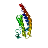
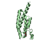
| Title | Crystal structure of Vps4-Vps20 complex from S.cerevisiae | ||||||
 Components Components |
| ||||||
 Keywords Keywords | VIRAL PROTEIN / MIT DOMAIN / ATPASE / YEAST | ||||||
| Function / homology |  Function and homology information Function and homology informationEndosomal Sorting Complex Required For Transport (ESCRT) / ESCRT IV complex / Sealing of the nuclear envelope (NE) by ESCRT-III / late endosome to lysosome transport via multivesicular body sorting pathway / intralumenal vesicle formation / Macroautophagy / : / protein retention in Golgi apparatus / ESCRT III complex / late endosome to vacuole transport via multivesicular body sorting pathway ...Endosomal Sorting Complex Required For Transport (ESCRT) / ESCRT IV complex / Sealing of the nuclear envelope (NE) by ESCRT-III / late endosome to lysosome transport via multivesicular body sorting pathway / intralumenal vesicle formation / Macroautophagy / : / protein retention in Golgi apparatus / ESCRT III complex / late endosome to vacuole transport via multivesicular body sorting pathway / sterol metabolic process / ATP export / nuclear membrane reassembly / vacuole organization / multivesicular body sorting pathway / midbody abscission / membrane fission / vesicle budding from membrane / plasma membrane repair / multivesicular body assembly / late endosome to vacuole transport / ubiquitin-dependent protein catabolic process via the multivesicular body sorting pathway / nucleus organization / reticulophagy / vacuolar membrane / endosomal transport / autophagosome maturation / multivesicular body / macroautophagy / autophagy / protein transport / midbody / endosome / endoplasmic reticulum / protein homodimerization activity / ATP hydrolysis activity / ATP binding / membrane / identical protein binding / plasma membrane / cytosol / cytoplasm Similarity search - Function | ||||||
| Biological species |  | ||||||
| Method |  X-RAY DIFFRACTION / SYNCHROTRON / MOLECULAR REPLACEMENT / Resolution: 1.973 Å X-RAY DIFFRACTION / SYNCHROTRON / MOLECULAR REPLACEMENT / Resolution: 1.973 Å | ||||||
 Authors Authors | Kojima, R. / Obita, T. / Onoue, K. / Mizuguchi, M. | ||||||
 Citation Citation | Journal: J.Mol.Biol. / Year: 2016 Title: Structural Fine-Tuning of Mit Interacting Motif 2 (Mim2) and Allosteric Regulation of Escrt-III by Vps4 in Yeast. Authors: Kojima, R. / Obita, T. / Onoue, K. / Mizuguchi, M. | ||||||
| History |
|
- Structure visualization
| Structure viewer | Molecule: MolmilJmol/JSmol |
|---|
- Downloads & links
-Download
| PDBx/mmCIF format | 5fvl.cif.gz | 51.5 KB | Display | PDBx/mmCIF format |
|---|---|---|---|---|
| PDB format | pdb5fvl.ent.gz | 38.2 KB | Display | PDB format |
| PDBx/mmJSON format | 5fvl.json.gz | Tree view | PDBx/mmJSON format | |
| Others |  Other downloads Other downloads |
-Validation report
| Arichive directory | https://data.pdbj.org/pub/pdb/validation_reports/fv/5fvlftp://data.pdbj.org/pub/pdb/validation_reports/fv/5fvl | HTTPS FTP |
|---|
-Related structure data
| Related structure data |  5fvkC 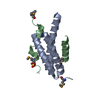 2v6xS C: citing same article ( S: Starting model for refinement |
|---|---|
| Similar structure data |


-Links
 PDBj
PDBj


- Assembly
| Deposited unit | 
| ||||||||
|---|---|---|---|---|---|---|---|---|---|
| 1 | 
| ||||||||
| 2 | 
| ||||||||
| Unit cell |
|
-Components
| #1: Protein | Mass: 9578.771 Da / Num. of mol.: 2 / Fragment: MIT DOMAIN, RESIDUES 1-82 Source method: isolated from a genetically manipulated source Source: (gene. exp.) Production host:  #2: Protein/peptide | Mass: 2887.267 Da / Num. of mol.: 2 / Fragment: RESIDUES 170-195 Source method: isolated from a genetically manipulated source Source: (gene. exp.) Production host: #3: Water | ChemComp-HOH / |  Mass: 18.015 Da / Num. of mol.: 25 / Source method: isolated from a natural source / Formula: H2O Mass: 18.015 Da / Num. of mol.: 25 / Source method: isolated from a natural source / Formula: H2O |
|---|
-Experimental details
-Experiment
| Experiment | Method: X-RAY DIFFRACTION / Number of used crystals: 1 |
|---|
- Sample preparation
| Crystal | Density Matthews: 1.83 Å3/Da / Density % sol: 32.62 % / Description: NONE |
|---|---|
| Crystal grow | pH: 8.6 / Details: pH 8.6 |
-Data collection
| Diffraction | Mean temperature: 100 K |
|---|---|
| Diffraction source | Source: SYNCHROTRON / Site: SPring-8  / Beamline: BL41XU / Wavelength: 1 / Beamline: BL41XU / Wavelength: 1 |
| Detector | Detector: CCD / Date: Dec 7, 2011 |
| Radiation | Protocol: SINGLE WAVELENGTH / Monochromatic (M) / Laue (L): M / Scattering type: x-ray |
| Radiation wavelength | Wavelength: 1 Å / Relative weight: 1 |
| Reflection | Resolution: 1.97→39.7 Å / Num. obs: 13410 / % possible obs: 100 % / Observed criterion σ(I): 2 / Redundancy: 14.3 % / Biso Wilson estimate: 16.94 Å2 / Rmerge(I) obs: 0.16 / Net I/σ(I): 15 |
| Reflection shell | Resolution: 1.97→2.08 Å / Redundancy: 14.3 % / Rmerge(I) obs: 0.52 / Mean I/σ(I) obs: 6.1 / % possible all: 100 |
- Processing
| Software |
| ||||||||||||||||||||||||||||||||||||||||||
|---|---|---|---|---|---|---|---|---|---|---|---|---|---|---|---|---|---|---|---|---|---|---|---|---|---|---|---|---|---|---|---|---|---|---|---|---|---|---|---|---|---|---|---|
| Refinement | Method to determine structure: MOLECULAR REPLACEMENT Starting model: PDB ENTRY 2V6X Resolution: 1.973→39.696 Å / SU ML: 0.19 / σ(F): 1.38 / Phase error: 20.08 / Stereochemistry target values: ML
| ||||||||||||||||||||||||||||||||||||||||||
| Solvent computation | Shrinkage radii: 0.9 Å / VDW probe radii: 1.11 Å / Solvent model: FLAT BULK SOLVENT MODEL | ||||||||||||||||||||||||||||||||||||||||||
| Refinement step | Cycle: LAST / Resolution: 1.973→39.696 Å
| ||||||||||||||||||||||||||||||||||||||||||
| Refine LS restraints |
| ||||||||||||||||||||||||||||||||||||||||||
| LS refinement shell |
|
