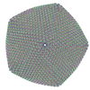
+ Open data
- Basic information
| Entry | Database: PDB / ID: 5j7v | ||||||
|---|---|---|---|---|---|---|---|
| Title | Faustovirus major capsid protein | ||||||
 Components Components | major capsid protein | ||||||
 Keywords Keywords | VIRUS / double jelly-roll | ||||||
| Function / homology | Group II dsDNA virus coat/capsid protein / Putative major capsid protein p72 Function and homology information Function and homology information | ||||||
| Biological species |  Faustovirus Faustovirus | ||||||
| Method | ELECTRON MICROSCOPY / single particle reconstruction / cryo EM / Resolution: 15.5 Å | ||||||
 Authors Authors | Klose, T. / Rossmann, M.G. | ||||||
| Funding support |  United States, 1items United States, 1items
| ||||||
 Citation Citation |  Journal: Proc Natl Acad Sci U S A / Year: 2016 Journal: Proc Natl Acad Sci U S A / Year: 2016Title: Structure of faustovirus, a large dsDNA virus. Authors: Thomas Klose / Dorine G Reteno / Samia Benamar / Adam Hollerbach / Philippe Colson / Bernard La Scola / Michael G Rossmann /   Abstract: Many viruses protect their genome with a combination of a protein shell with or without a membrane layer. Here we describe the structure of faustovirus, the first DNA virus (to our knowledge) that ...Many viruses protect their genome with a combination of a protein shell with or without a membrane layer. Here we describe the structure of faustovirus, the first DNA virus (to our knowledge) that has been found to use two protein shells to encapsidate and protect its genome. The crystal structure of the major capsid protein, in combination with cryo-electron microscopy structures of two different maturation stages of the virus, shows that the outer virus shell is composed of a double jelly-roll protein that can be found in many double-stranded DNA viruses. The structure of the repeating hexameric unit of the inner shell is different from all other known capsid proteins. In addition to the unique architecture, the region of the genome that encodes the major capsid protein stretches over 17,000 bp and contains a large number of introns and exons. This complexity might help the virus to rapidly adapt to new environments or hosts. | ||||||
| History |
|
- Structure visualization
| Movie |
 Movie viewer Movie viewer |
|---|---|
| Structure viewer | Molecule:  Molmil Molmil Jmol/JSmol Jmol/JSmol |
- Downloads & links
- Download
| PDBx/mmCIF format |  5j7v.cif.gz 5j7v.cif.gz | 505.6 KB | Display |  PDBx/mmCIF format PDBx/mmCIF format |
|---|---|---|---|---|
| PDB format |  pdb5j7v.ent.gz pdb5j7v.ent.gz | Display |  PDB format PDB format | |
| PDBx/mmJSON format |  5j7v.json.gz 5j7v.json.gz | Tree view |  PDBx/mmJSON format PDBx/mmJSON format | |
| Others |  Other downloads Other downloads |
-Validation report
| Summary document |  5j7v_validation.pdf.gz 5j7v_validation.pdf.gz | 1 MB | Display |  wwPDB validaton report wwPDB validaton report |
|---|---|---|---|---|
| Full document |  5j7v_full_validation.pdf.gz 5j7v_full_validation.pdf.gz | 1.1 MB | Display | |
| Data in XML |  5j7v_validation.xml.gz 5j7v_validation.xml.gz | 57.6 KB | Display | |
| Data in CIF |  5j7v_validation.cif.gz 5j7v_validation.cif.gz | 87.4 KB | Display | |
| Arichive directory |  https://data.pdbj.org/pub/pdb/validation_reports/j7/5j7v https://data.pdbj.org/pub/pdb/validation_reports/j7/5j7v ftp://data.pdbj.org/pub/pdb/validation_reports/j7/5j7v ftp://data.pdbj.org/pub/pdb/validation_reports/j7/5j7v | HTTPS FTP |
-Related structure data
| Related structure data |  8144MC  8145C  5j7oC  5j7uC M: map data used to model this data C: citing same article ( |
|---|---|
| Similar structure data |
- Links
Links
- Assembly
| Deposited unit | 
|
|---|---|
| 1 | x 2760
|
| 2 | x 46
|
| 3 | x 230
|
| 4 | x 46
|
| Symmetry | Point symmetry: (Schoenflies symbol: I (icosahedral)) |
- Components
| #1: Protein | Mass: 71781.891 Da / Num. of mol.: 3 / Source method: isolated from a natural source / Source: (natural)  Faustovirus / References: UniProt: A0A0H3TLP8*PLUS Faustovirus / References: UniProt: A0A0H3TLP8*PLUS |
|---|
-Experimental details
-Experiment
| Experiment | Method: ELECTRON MICROSCOPY |
|---|---|
| EM experiment | Aggregation state: PARTICLE / 3D reconstruction method: single particle reconstruction |
- Sample preparation
| Component | Name: Faustovirus / Type: VIRUS / Entity ID: all / Source: NATURAL | |||||||||||||||
|---|---|---|---|---|---|---|---|---|---|---|---|---|---|---|---|---|
| Source (natural) | Organism:  Faustovirus Faustovirus | |||||||||||||||
| Details of virus | Empty: NO / Enveloped: NO / Isolate: SPECIES / Type: VIRION | |||||||||||||||
| Natural host | Organism: Vermamoeba vermiformis / Strain: CDC 19 | |||||||||||||||
| Virus shell |
| |||||||||||||||
| Buffer solution | pH: 7 | |||||||||||||||
| Specimen | Embedding applied: NO / Shadowing applied: NO / Staining applied: NO / Vitrification applied: YES | |||||||||||||||
| Vitrification | Instrument: GATAN CRYOPLUNGE 3 / Cryogen name: ETHANE / Details: Plunged into liquid ethane |
- Electron microscopy imaging
| Experimental equipment |  Model: Titan Krios / Image courtesy: FEI Company |
|---|---|
| Microscopy | Model: FEI TITAN KRIOS |
| Electron gun | Electron source:  FIELD EMISSION GUN / Accelerating voltage: 300 kV / Illumination mode: SPOT SCAN FIELD EMISSION GUN / Accelerating voltage: 300 kV / Illumination mode: SPOT SCAN |
| Electron lens | Mode: BRIGHT FIELD / Cs: 2.7 mm / C2 aperture diameter: 100 µm / Alignment procedure: COMA FREE |
| Specimen holder | Cryogen: NITROGEN / Specimen holder model: FEI TITAN KRIOS AUTOGRID HOLDER |
| Image recording | Electron dose: 25 e/Å2 / Film or detector model: GATAN ULTRASCAN 4000 (4k x 4k) |
- Processing
| EM software |
| ||||||||||||||||||||||||||||||||||||||||
|---|---|---|---|---|---|---|---|---|---|---|---|---|---|---|---|---|---|---|---|---|---|---|---|---|---|---|---|---|---|---|---|---|---|---|---|---|---|---|---|---|---|
| CTF correction | Type: PHASE FLIPPING ONLY | ||||||||||||||||||||||||||||||||||||||||
| Symmetry | Point symmetry: I (icosahedral) | ||||||||||||||||||||||||||||||||||||||||
| 3D reconstruction | Resolution: 15.5 Å / Resolution method: FSC 0.143 CUT-OFF / Num. of particles: 9640 / Algorithm: FOURIER SPACE / Symmetry type: POINT | ||||||||||||||||||||||||||||||||||||||||
| Atomic model building | Protocol: RIGID BODY FIT / Space: REAL / Target criteria: Correlation coefficient | ||||||||||||||||||||||||||||||||||||||||
| Atomic model building | 3D fitting-ID: 1 / Accession code: 5J7O / Initial refinement model-ID: 1 / PDB-ID: 5J7O / Source name: PDB / Type: experimental model
|
 Movie
Movie Controller


 PDBj
PDBj

