 Movie
Movie Controller

[English] 日本語
 Yorodumi
- PDB-4v93: Fitted coordinates for Lumbricus terrestris hemoglobin cryo-EM co... -
+ Open data

- Basic information
| Entry | Database: PDB / ID: 4v93 | |||||||||
|---|---|---|---|---|---|---|---|---|---|---|
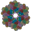
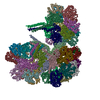
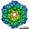
| Title | Fitted coordinates for Lumbricus terrestris hemoglobin cryo-EM complex (EMD-2627) | |||||||||
 Components Components |
| |||||||||
 Keywords Keywords | TRANSPORT PROTEIN | |||||||||
| Function / homology |  Function and homology information Function and homology informationhemoglobin complex / oxygen carrier activity / oxygen binding / response to hypoxia / iron ion binding / heme binding / extracellular region / metal ion binding Similarity search - Function | |||||||||
| Biological species |  LUMBRICUS TERRESTRIS (common earthworm) LUMBRICUS TERRESTRIS (common earthworm) | |||||||||
| Method | ELECTRON MICROSCOPY / single particle reconstruction / cryo EM / Resolution: 8.1 Å | |||||||||
 Authors Authors | Chen, W.T. / Chen, Y.C. / Liou, H.H. / Chao, C.Y. | |||||||||
 Citation Citation | Journal: Sci Rep / Year: 2015 Title: Structural basis for cooperative oxygen binding and bracelet-assisted assembly of Lumbricus terrestris hemoglobin. Authors: Wei-Ting Chen / Yu-Chuen Chen / Horng-Huei Liou / Chih-Yu Chao /  Abstract: The iron-containing hemoglobins (Hbs) are essential proteins to serve as oxygen transporters in the blood. Among various kinds of Hbs, the earthworm Hbs are the champions in carrying oxygen due to ...The iron-containing hemoglobins (Hbs) are essential proteins to serve as oxygen transporters in the blood. Among various kinds of Hbs, the earthworm Hbs are the champions in carrying oxygen due to not only their large size but also the unusually high cooperativity of ligand binding. However, the cooperative oxygen binding mechanisms are still mostly unknown. Here we report the cryo-electron microscopy structure of Lumbricus terrestris Hb in its native, oxygenated state at 9.1 Å resolution, showing remarkable differences from the carbon monoxide-binding X-ray structure. Our structural analysis first indicates that the cooperative ligand binding of L. terrestris Hb requires tertiary and quaternary transitions in the heme pocket and a global subunit movement facilitated by intra-ring and inter-ring contacts. Moreover, the additional sinusoidal bracelet provides the confirmation for the long-standing debate about the additional electron densities absent in the X-ray crystal structure. | |||||||||
| History |
|
- Structure visualization
| Movie |
Movie viewer |
|---|---|
| Structure viewer | Molecule: MolmilJmol/JSmol |
- Downloads & links
-Download
| PDBx/mmCIF format | 4v93.cif.gz | 4.9 MB | Display | PDBx/mmCIF format |
|---|---|---|---|---|
| PDB format | pdb4v93.ent.gz | Display | PDB format | |
| PDBx/mmJSON format | 4v93.json.gz | Tree view | PDBx/mmJSON format | |
| Others |  Other downloads Other downloads |
-Validation report
| Summary document | 4v93_validation.pdf.gz | 1.4 MB | Display | wwPDB validaton report |
|---|---|---|---|---|
| Full document | 4v93_full_validation.pdf.gz | 1.8 MB | Display | |
| Data in XML | 4v93_validation.xml.gz | 641.4 KB | Display | |
| Data in CIF | 4v93_validation.cif.gz | 1012.7 KB | Display | |
| Arichive directory | https://data.pdbj.org/pub/pdb/validation_reports/v9/4v93ftp://data.pdbj.org/pub/pdb/validation_reports/v9/4v93 | HTTPS FTP |
-Related structure data
| Related structure data |  2627MC M: map data used to model this data C: citing same article ( |
|---|---|
| Similar structure data |


-Links
 PDBj
PDBj


- Assembly
| Deposited unit | 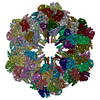
|
|---|---|
| 1 |
|
-Components
-EXTRACELLULAR GLOBIN- ... , 5 types, 108 molecules A0A5ACAHAMARAWAbAgAlAqAvA2A3A7AAAEAFAJAKAOAPATAUAYAZAdAeAiAj...
| #1: Protein | Mass: 16902.410 Da / Num. of mol.: 12 / Source method: isolated from a natural source / Source: (natural) LUMBRICUS TERRESTRIS (common earthworm) / References: UniProt: P11069#3: Protein | Mass: 17136.619 Da / Num. of mol.: 24 / Source method: isolated from a natural source / Source: (natural) LUMBRICUS TERRESTRIS (common earthworm) / References: UniProt: P13579#4: Protein | Mass: 16268.229 Da / Num. of mol.: 36 / Source method: isolated from a natural source / Source: (natural) LUMBRICUS TERRESTRIS (common earthworm) / References: UniProt: P02218#6: Protein | Mass: 17566.990 Da / Num. of mol.: 12 / Source method: isolated from a natural source / Source: (natural) LUMBRICUS TERRESTRIS (common earthworm) / References: UniProt: P13579#7: Protein | Mass: 19126.137 Da / Num. of mol.: 24 / Source method: isolated from a natural source / Source: (natural) LUMBRICUS TERRESTRIS (common earthworm) / References: UniProt: P11069 |
|---|
-HEMOGLOBIN CHAIN ... , 2 types, 36 molecules A1A6ADAIANASAXAcAhAmArAwB0B5BCBHBMBRBWBbBgBlBqBvC4CBCGCLCQCV...
| #2: Protein | Mass: 15988.263 Da / Num. of mol.: 12 / Source method: isolated from a natural source / Source: (natural) LUMBRICUS TERRESTRIS (common earthworm) / References: UniProt: O61233#5: Protein | Mass: 17948.670 Da / Num. of mol.: 24 / Source method: isolated from a natural source / Source: (natural) LUMBRICUS TERRESTRIS (common earthworm) / References: UniProt: O61233 |
|---|
-Protein , 1 types, 12 molecules C0C5CCCHCMCRCWCbCgClCqCv
| #8: Protein | Mass: 27460.688 Da / Num. of mol.: 12 / Source method: isolated from a natural source / Source: (natural) LUMBRICUS TERRESTRIS (common earthworm) / References: UniProt: Q9GV76 |
|---|
-EXTRACELLULAR HEMOGLOBIN LINKER ... , 2 types, 24 molecules C1C6CDCICNCSCXCcChCmCrCwC2C7CECJCOCTCYCdCiCnCsCx
| #9: Protein | Mass: 32075.490 Da / Num. of mol.: 12 / Source method: isolated from a natural source / Source: (natural) LUMBRICUS TERRESTRIS (common earthworm) / References: UniProt: Q2I743#10: Protein | Mass: 26877.914 Da / Num. of mol.: 12 / Source method: isolated from a natural source / Source: (natural) LUMBRICUS TERRESTRIS (common earthworm) / References: UniProt: Q2I742 |
|---|
-Experimental details
-Experiment
| Experiment | Method: ELECTRON MICROSCOPY |
|---|---|
| EM experiment | Aggregation state: PARTICLE / 3D reconstruction method: single particle reconstruction |
- Sample preparation
| Component | Name: Lumbricus terrestris hemoglobin / Type: COMPLEX |
|---|---|
| Buffer solution | pH: 7.2 |
| Specimen | Embedding applied: NO / Shadowing applied: NO / Staining applied: NO / Vitrification applied: YES |
| Specimen support | Details: HOLEY CARBON |
| Vitrification | Instrument: FEI VITROBOT MARK III / Cryogen name: ETHANE |
- Electron microscopy imaging
| Experimental equipment |  Model: Tecnai F20 / Image courtesy: FEI Company |
|---|---|
| Microscopy | Model: FEI TECNAI F20 / Date: Oct 15, 2012 |
| Electron gun | Electron source:  FIELD EMISSION GUN / Accelerating voltage: 200 kV / Illumination mode: FLOOD BEAM FIELD EMISSION GUN / Accelerating voltage: 200 kV / Illumination mode: FLOOD BEAM |
| Electron lens | Mode: BRIGHT FIELD / Nominal magnification: 80000 X |
| Image recording | Film or detector model: GATAN ULTRASCAN 4000 (4k x 4k) |
| Radiation wavelength | Relative weight: 1 |
- Processing
| Symmetry | Point symmetry: D6 (2x6 fold dihedral) | ||||||||||||
|---|---|---|---|---|---|---|---|---|---|---|---|---|---|
| 3D reconstruction | Resolution: 8.1 Å / Num. of particles: 4500 Details: SUBMISSION BASED ON EXPERIMENTAL DATA FROM EMDB EMD -2627. (DEPOSITION ID: 12397). Symmetry type: POINT | ||||||||||||
| Atomic model building | Protocol: OTHER / Details: REFINEMENT PROTOCOL--X-RAY | ||||||||||||
| Atomic model building | PDB-ID: 2GTL | ||||||||||||
| Refinement | Highest resolution: 8.1 Å | ||||||||||||
| Refinement step | Cycle: LAST / Highest resolution: 8.1 Å
|

