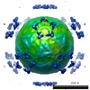
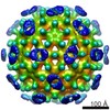
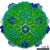
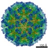
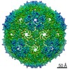
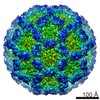
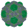
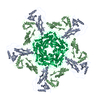
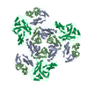
- PDB-4pt2: Myxococcus xanthus encapsulin protein (EncA) -
+
データを開く
IDまたはキーワード:
読み込み中...
-
基本情報
登録情報
データベース: PDB / ID: 4pt2
タイトル
Myxococcus xanthus encapsulin protein (EncA)
要素
Encapsulin protein
キーワード
VIRUS LIKE PARTICLE / HK97 fold / Shell protein
機能・相同性
Type 1 encapsulin shell protein / Encapsulating protein for peroxidase / encapsulin nanocompartment / iron ion transport / intracellular iron ion homeostasis / Type 1 encapsulin shell protein EncA
ジャーナル: EMBO J / 年: 2014 タイトル: A virus capsid-like nanocompartment that stores iron and protects bacteria from oxidative stress. 著者: Colleen A McHugh / Juan Fontana / Daniel Nemecek / Naiqian Cheng / Anastasia A Aksyuk / J Bernard Heymann / Dennis C Winkler / Alan S Lam / Joseph S Wall / Alasdair C Steven / Egbert Hoiczyk / 要旨: Living cells compartmentalize materials and enzymatic reactions to increase metabolic efficiency. While eukaryotes use membrane-bound organelles, bacteria and archaea rely primarily on protein-bound ...Living cells compartmentalize materials and enzymatic reactions to increase metabolic efficiency. While eukaryotes use membrane-bound organelles, bacteria and archaea rely primarily on protein-bound nanocompartments. Encapsulins constitute a class of nanocompartments widespread in bacteria and archaea whose functions have hitherto been unclear. Here, we characterize the encapsulin nanocompartment from Myxococcus xanthus, which consists of a shell protein (EncA, 32.5 kDa) and three internal proteins (EncB, 17 kDa; EncC, 13 kDa; EncD, 11 kDa). Using cryo-electron microscopy, we determined that EncA self-assembles into an icosahedral shell 32 nm in diameter (26 nm internal diameter), built from 180 subunits with the fold first observed in bacteriophage HK97 capsid. The internal proteins, of which EncB and EncC have ferritin-like domains, attach to its inner surface. Native nanocompartments have dense iron-rich cores. Functionally, they resemble ferritins, cage-like iron storage proteins, but with a massively greater capacity (~30,000 iron atoms versus ~3,000 in ferritin). Physiological data reveal that few nanocompartments are assembled during vegetative growth, but they increase fivefold upon starvation, protecting cells from oxidative stress through iron sequestration.
履歴
登録
2014年3月10日
登録サイト: RCSB / 処理サイト: RCSB
改定 1.0
2014年7月30日
Provider: repository / タイプ: Initial release
改定 1.1
2014年9月10日
Group: Database references
改定 1.2
2018年7月18日
Group: Data collection / カテゴリ: em_software / Item: _em_software.image_processing_id
手法: reference based / 解像度: 4.6 Å / 解像度の算出法: FSC 0.143 CUT-OFF / 粒子像の数: 14000 / ピクセルサイズ(公称値): 1.102 Å / ピクセルサイズ(実測値): 1.102 Å 詳細: Final map was calculated dividing particles into two independent data sets 対称性のタイプ: POINT
精密化
解像度: 4.6→771.4 Å / SU ML: 1.04 / σ(F): 0 / 位相誤差: 43.52 / 立体化学のターゲット値: MLHL 詳細: Homology model of PDB entry 3DKT (I-TASSER), flexible fitting using MDFF and manual re-building using COOT, refined using structure factors derived from EMD-5917
Rfactor
反射数
%反射
Rwork
0.3489
-
-
obs
0.3489
9859810
99.83 %
all
-
9859810
-
溶媒の処理
減衰半径: 0.9 Å / VDWプローブ半径: 1.11 Å / 溶媒モデル: FLAT BULK SOLVENT MODEL
 ムービー
ムービー コントローラー


 データを開く
 基本情報
基本情報 要素
要素 キーワード
キーワード 機能・相同性情報
 Myxococcus xanthus (バクテリア)
 データ登録者
 引用
引用
 構造の表示
構造の表示 ダウンロードとリンク
 その他のダウンロード
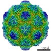


 PDBj
PDBj
 集合体
集合体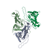

 試料調製
試料調製 電子顕微鏡撮影
 FIELD EMISSION GUN / 加速電圧: 300 kV / 照射モード: FLOOD BEAM
 解析
解析