+ データを開く
- 基本情報
基本情報
| 登録情報 | データベース: PDB / ID: 4d5n | |||||||||
|---|---|---|---|---|---|---|---|---|---|---|
| タイトル | Cryo-EM structures of ribosomal 80S complexes with termination factors and cricket paralysis virus IRES reveal the IRES in the translocated state | |||||||||
 要素 要素 |
| |||||||||
 キーワード キーワード | RIBOSOME/RNA / RIBOSOME-RNA COMPLEX / CRPV IRES / RIBOSOME / TERMINATION / RELEASE FACTORS | |||||||||
| 機能・相同性 |  機能・相同性情報 機能・相同性情報translation termination factor activity / translation release factor complex / cytoplasmic translational termination / regulation of translational termination / translation release factor activity, codon specific / protein methylation / translation release factor activity / sequence-specific mRNA binding / peptidyl-tRNA hydrolase activity / nuclear-transcribed mRNA catabolic process, nonsense-mediated decay ...translation termination factor activity / translation release factor complex / cytoplasmic translational termination / regulation of translational termination / translation release factor activity, codon specific / protein methylation / translation release factor activity / sequence-specific mRNA binding / peptidyl-tRNA hydrolase activity / nuclear-transcribed mRNA catabolic process, nonsense-mediated decay / Protein hydroxylation / Eukaryotic Translation Termination / Nonsense Mediated Decay (NMD) independent of the Exon Junction Complex (EJC) / Nonsense Mediated Decay (NMD) enhanced by the Exon Junction Complex (EJC) / translational termination / cytosolic ribosome / Regulation of expression of SLITs and ROBOs / ribosome binding / RNA binding / cytosol / cytoplasm 類似検索 - 分子機能 | |||||||||
| 生物種 |  HOMO SAPIENS (ヒト) HOMO SAPIENS (ヒト) CRICKET PARALYSIS VIRUS (ウイルス) CRICKET PARALYSIS VIRUS (ウイルス) | |||||||||
| 手法 | 電子顕微鏡法 / 単粒子再構成法 / クライオ電子顕微鏡法 / 解像度: 9 Å | |||||||||
 データ登録者 データ登録者 | Muhs, M. / Hilal, T. / Mielke, T. / Skabkin, M.A. / Sanbonmatsu, K.Y. / Pestova, T.V. / Spahn, C.M.T. | |||||||||
 引用 引用 |  ジャーナル: Mol Cell / 年: 2015 ジャーナル: Mol Cell / 年: 2015タイトル: Cryo-EM of ribosomal 80S complexes with termination factors reveals the translocated cricket paralysis virus IRES. 著者: Margarita Muhs / Tarek Hilal / Thorsten Mielke / Maxim A Skabkin / Karissa Y Sanbonmatsu / Tatyana V Pestova / Christian M T Spahn /   要旨: The cricket paralysis virus (CrPV) uses an internal ribosomal entry site (IRES) to hijack the ribosome. In a remarkable RNA-based mechanism involving neither initiation factor nor initiator tRNA, the ...The cricket paralysis virus (CrPV) uses an internal ribosomal entry site (IRES) to hijack the ribosome. In a remarkable RNA-based mechanism involving neither initiation factor nor initiator tRNA, the CrPV IRES jumpstarts translation in the elongation phase from the ribosomal A site. Here, we present cryoelectron microscopy (cryo-EM) maps of 80S⋅CrPV-STOP ⋅ eRF1 ⋅ eRF3 ⋅ GMPPNP and 80S⋅CrPV-STOP ⋅ eRF1 complexes, revealing a previously unseen binding state of the IRES and directly rationalizing that an eEF2-dependent translocation of the IRES is required to allow the first A-site occupation. During this unusual translocation event, the IRES undergoes a pronounced conformational change to a more stretched conformation. At the same time, our structural analysis provides information about the binding modes of eRF1 ⋅ eRF3 ⋅ GMPPNP and eRF1 in a minimal system. It shows that neither eRF3 nor ABCE1 are required for the active conformation of eRF1 at the intersection between eukaryotic termination and recycling. | |||||||||
| 履歴 |
|
- 構造の表示
構造の表示
| ムービー |
 ムービービューア ムービービューア |
|---|---|
| 構造ビューア | 分子:  Molmil Molmil Jmol/JSmol Jmol/JSmol |
- ダウンロードとリンク
- ダウンロード
| PDBx/mmCIF形式 |  4d5n.cif.gz 4d5n.cif.gz | 230 KB | 表示 |  PDBx/mmCIF形式 PDBx/mmCIF形式 |
|---|---|---|---|---|
| PDB形式 |  pdb4d5n.ent.gz pdb4d5n.ent.gz | 169.8 KB | 表示 |  PDB形式 PDB形式 |
| PDBx/mmJSON形式 |  4d5n.json.gz 4d5n.json.gz | ツリー表示 |  PDBx/mmJSON形式 PDBx/mmJSON形式 | |
| その他 |  その他のダウンロード その他のダウンロード |
-検証レポート
| 文書・要旨 |  4d5n_validation.pdf.gz 4d5n_validation.pdf.gz | 799.6 KB | 表示 |  wwPDB検証レポート wwPDB検証レポート |
|---|---|---|---|---|
| 文書・詳細版 |  4d5n_full_validation.pdf.gz 4d5n_full_validation.pdf.gz | 860.1 KB | 表示 | |
| XML形式データ |  4d5n_validation.xml.gz 4d5n_validation.xml.gz | 30.6 KB | 表示 | |
| CIF形式データ |  4d5n_validation.cif.gz 4d5n_validation.cif.gz | 44.9 KB | 表示 | |
| アーカイブディレクトリ |  https://data.pdbj.org/pub/pdb/validation_reports/d5/4d5n https://data.pdbj.org/pub/pdb/validation_reports/d5/4d5n ftp://data.pdbj.org/pub/pdb/validation_reports/d5/4d5n ftp://data.pdbj.org/pub/pdb/validation_reports/d5/4d5n | HTTPS FTP |
-関連構造データ
| 関連構造データ |  2810MC  2813C  4d5lC  4d5yC  4d61C  4d67C  4d66  4d68 C: 同じ文献を引用 ( M: このデータのモデリングに利用したマップデータ |
|---|---|
| 類似構造データ |
- リンク
リンク
- 集合体
集合体
| 登録構造単位 | 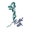
|
|---|---|
| 1 |
|
- 要素
要素
| #1: タンパク質 | 分子量: 49040.711 Da / 分子数: 1 / 断片: RESIDUES 5-437 / 由来タイプ: 組換発現 / 由来: (組換発現)  HOMO SAPIENS (ヒト) / 発現宿主: HOMO SAPIENS (ヒト) / 発現宿主:  |
|---|---|
| #2: RNA鎖 | 分子量: 64363.910 Da / 分子数: 1 / 由来タイプ: 合成 / 由来: (合成)  CRICKET PARALYSIS VIRUS (ウイルス) / 参照: GenBank: 8895506 CRICKET PARALYSIS VIRUS (ウイルス) / 参照: GenBank: 8895506 |
| 配列の詳細 | FIRST CODING TRIPLET MUTATED TO STOP |
-実験情報
-実験
| 実験 | 手法: 電子顕微鏡法 |
|---|---|
| EM実験 | 試料の集合状態: PARTICLE / 3次元再構成法: 単粒子再構成法 |
- 試料調製
試料調製
| 構成要素 | 名称: CRICKET PARALYSIS VIRUS IRES RNA BOUND TO MAMMALIAN 80S RIBOSOME AND ERF1 タイプ: RIBOSOME / 詳細: MICROGRAPHS SELECTED FOR ASTIGMATISM AND DRIFT |
|---|---|
| 緩衝液 | 名称: 20 MM TRIS PH 7.5, 100 MM KCL, 1 MM DTT, 2.5 MM MGCL2, 0.5 MM GTP pH: 7.5 詳細: 20 MM TRIS PH 7.5, 100 MM KCL, 1 MM DTT, 2.5 MM MGCL2, 0.5 MM GTP |
| 試料 | 濃度: 1.38 mg/ml / 包埋: NO / シャドウイング: NO / 染色: NO / 凍結: YES |
| 試料支持 | 詳細: HOLEY CARBON |
| 急速凍結 | 装置: FEI VITROBOT MARK II / 凍結剤: ETHANE / 詳細: LIQUID ETHANE |
- 電子顕微鏡撮影
| 実験機器 |  モデル: Tecnai F20 / 画像提供: FEI Company |
|---|---|
| 顕微鏡 | モデル: FEI TECNAI F20 / 日付: 2012年4月17日 / 詳細: GOOD MICROGRAPHS SELECTED FOR ASTIGMATISM AND DRIFT |
| 電子銃 | 電子線源:  FIELD EMISSION GUN / 加速電圧: 300 kV / 照射モード: FLOOD BEAM FIELD EMISSION GUN / 加速電圧: 300 kV / 照射モード: FLOOD BEAM |
| 電子レンズ | モード: BRIGHT FIELD / 倍率(公称値): 39000 X / 倍率(補正後): 65520 X / 最大 デフォーカス(公称値): 4000 nm / 最小 デフォーカス(公称値): 2000 nm / Cs: 2 mm |
| 試料ホルダ | 温度: 77 K |
| 撮影 | 電子線照射量: 20 e/Å2 / フィルム・検出器のモデル: KODAK SO-163 FILM |
| 画像スキャン | デジタル画像の数: 366 |
- 解析
解析
| EMソフトウェア |
| ||||||||||||
|---|---|---|---|---|---|---|---|---|---|---|---|---|---|
| CTF補正 | 詳細: DEFOCUS GROUPS | ||||||||||||
| 対称性 | 点対称性: C1 (非対称) | ||||||||||||
| 3次元再構成 | 手法: MULTI-REFERENCE TEMPLATE MATCHING / 解像度: 9 Å / 粒子像の数: 109596 / ピクセルサイズ(公称値): 1.56 Å / ピクセルサイズ(実測値): 1.56 Å 倍率補正: CROSS- -CORRELATION DENSITIES WITH REFERENCE STRUCTURE 詳細: SUBMISSION BASED ON EXPERIMENTAL DATA FROM EMDB EMD-2810. (DEPOSITION ID: 12907). 対称性のタイプ: POINT | ||||||||||||
| 原子モデル構築 | プロトコル: FLEXIBLE FIT / 空間: REAL / 詳細: METHOD--RIGID BODY, FLEXIBLE FIT | ||||||||||||
| 精密化 | 最高解像度: 9 Å | ||||||||||||
| 精密化ステップ | サイクル: LAST / 最高解像度: 9 Å
|
 ムービー
ムービー コントローラー



 PDBj
PDBj


