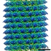
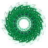
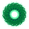
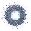
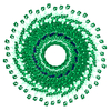
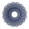
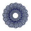
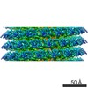
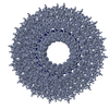
ジャーナル: Proc Natl Acad Sci U S A / 年: 2011 タイトル: Hydrogen-bonding networks and RNA bases revealed by cryo electron microscopy suggest a triggering mechanism for calcium switches. 著者: Peng Ge / Z Hong Zhou / 要旨: Helical assemblies such as filamentous viruses, flagella, and F-actin represent an important category of structures in biology. As the first discovered virus, tobacco mosaic virus (TMV) was at the ...Helical assemblies such as filamentous viruses, flagella, and F-actin represent an important category of structures in biology. As the first discovered virus, tobacco mosaic virus (TMV) was at the center of virus research. Previously, the structure of TMV was solved at atomic detail by X-ray fiber diffraction but only for its dormant or high-calcium-concentration state, not its low-calcium-concentration state, which is relevant to viral assembly and disassembly inside host cells. Here we report a helical reconstruction of TMV in its calcium-free, metastable assembling state at 3.3 Å resolution by cryo electron microscopy, revealing both protein side chains and RNA bases. An atomic model was built de novo showing marked differences from the high-calcium, dormant-state structure. Although it could be argued that there might be inaccuracies in the latter structure derived from X-ray fiber diffraction, these differences can be interpreted as conformational changes effected by calcium-driven switches, a common regulatory mechanism in plant viruses. Our comparisons of the structures of the low- and high-calcium states indicate that hydrogen bonds formed by Asp116 and Arg92 in the place of the calcium ion of the dormant (high-calcium) state might trigger allosteric changes in the RNA base-binding pockets of the coat protein. In turn, the coat protein-RNA interactions in our structure favor an adenine-X-guanine (A*G) motif over the G*A motif of the dormant state, thus offering an explanation underlying viral assembly initiation by an AAG motif.
名称: Tris-buffered saline / pH: 7.4 / 詳細: 10 mM Tris, 130 mM sodium chloride
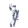
試料
包埋: NO / シャドウイング: NO / 染色: NO / 凍結: YES / 詳細: 10 mM Tris, 130 mM sodium chloride
試料支持
詳細: 400 mesh Quantifoil 1.3/1.2 micro-meter
急速凍結
装置: FEI VITROBOT MARK IV / 凍結剤: ETHANE / Temp: 93 K / 湿度: 100 % / 詳細: 2 uL sample 手法: 10 second wait time, 1-2 second blot time, 1-2 second drain time, blot force 1
-
電子顕微鏡撮影
実験機器
モデル: Titan Krios / 画像提供: FEI Company
顕微鏡
モデル: FEI TITAN KRIOS / 日付: 2009年6月19日
電子銃
電子線源: FIELD EMISSION GUN / 加速電圧: 300 kV / 照射モード: FLOOD BEAM / Electron beam tilt params: 0
電子レンズ
モード: BRIGHT FIELD / 倍率(公称値): 75000 X / 倍率(補正後): 73000 X / 最大 デフォーカス(公称値): 2500 nm / 最小 デフォーカス(公称値): 1200 nm / Cs: 2.7 mm 非点収差: objective lens astigmatism was corrected at 250000x magnification カメラ長: 0 mm
試料ホルダ
試料ホルダーモデル: SIDE ENTRY, EUCENTRIC / 資料ホルダタイプ: Eucentric / 温度: 90 K / 傾斜角・最大: 0 ° / 傾斜角・最小: 0 °
撮影
電子線照射量: 25 e/Å2 / フィルム・検出器のモデル: KODAK SO-163 FILM
画像スキャン
デジタル画像の数: 386
放射
プロトコル: SINGLE WAVELENGTH / 単色(M)・ラウエ(L): M / 散乱光タイプ: x-ray
放射波長
相対比: 1
-
解析
ソフトウェア
名称: EMAN / 分類: 精密化
EMソフトウェア
ID
名称
カテゴリ
1
EMAN
3次元再構成
2
IHRSR
3次元再構成
らせん対称
回転角度/サブユニット: 22.035 ° / 軸方向距離/サブユニット: 1.407 Å / らせん対称軸の対称性: C1
3次元再構成
手法: IHRSR / 解像度: 3.3 Å / 解像度の算出法: OTHER / ピクセルサイズ(公称値): 0.847 Å / ピクセルサイズ(実測値): 0.869 Å / 対称性のタイプ: HELICAL
 ムービー
ムービー コントローラー


 データを開く
 基本情報
基本情報 要素
要素 キーワード
キーワード 機能・相同性情報
 Tobacco mosaic virus (ウイルス)
 データ登録者
 引用
引用
 構造の表示
構造の表示 ダウンロードとリンク
 その他のダウンロード
 PDBj
PDBj
 集合体
集合体
 試料調製
試料調製 電子顕微鏡撮影
 FIELD EMISSION GUN / 加速電圧: 300 kV / 照射モード: FLOOD BEAM / Electron beam tilt params: 0
  解析
解析