 ムービー
ムービー コントローラー


+ データを開く

- 基本情報
基本情報
| 登録情報 | データベース: PDB / ID: 3g37 | ||||||
|---|---|---|---|---|---|---|---|
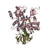
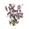
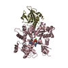
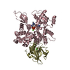
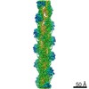
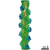
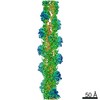
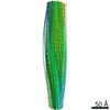
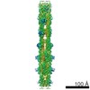
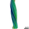
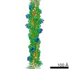
| タイトル | Cryo-EM structure of actin filament in the presence of phosphate | ||||||
 要素 要素 | Actin, alpha skeletal muscle | ||||||
 キーワード キーワード | CONTRACTILE PROTEIN / actin / cytoskeleton / cell adhesion / cellular signaling / cytokinesis / muscle / cryo-EM / ATP-binding / Methylation / Muscle protein / Nucleotide-binding / Phosphoprotein | ||||||
| 機能・相同性 |  機能・相同性情報 機能・相同性情報cytoskeletal motor activator activity / tropomyosin binding / mesenchyme migration / troponin I binding / myosin heavy chain binding / filamentous actin / actin filament bundle / skeletal muscle thin filament assembly / striated muscle thin filament / actin filament bundle assembly ...cytoskeletal motor activator activity / tropomyosin binding / mesenchyme migration / troponin I binding / myosin heavy chain binding / filamentous actin / actin filament bundle / skeletal muscle thin filament assembly / striated muscle thin filament / actin filament bundle assembly / skeletal muscle myofibril / actin monomer binding / skeletal muscle fiber development / stress fiber / titin binding / actin filament polymerization / filopodium / actin filament / 加水分解酵素; 酸無水物に作用; 酸無水物に作用・細胞または細胞小器官の運動に関与 / calcium-dependent protein binding / lamellipodium / cell body / hydrolase activity / protein domain specific binding / calcium ion binding / positive regulation of gene expression / magnesium ion binding / ATP binding / identical protein binding / cytoplasm 類似検索 - 分子機能 | ||||||
| 生物種 |  | ||||||
| 手法 | 電子顕微鏡法 / 単粒子再構成法 / クライオ電子顕微鏡法 / 解像度: 6 Å | ||||||
 データ登録者 データ登録者 | Wakabayshi, T. / Murakami, K. / Yasunaga, T. / Noguchi, T.Q. / Uyeda, T.Q. | ||||||
 引用 引用 | ジャーナル: Cell / 年: 2010 タイトル: Structural basis for actin assembly, activation of ATP hydrolysis, and delayed phosphate release. 著者: Kenji Murakami / Takuo Yasunaga / Taro Q P Noguchi / Yuki Gomibuchi / Kien X Ngo / Taro Q P Uyeda / Takeyuki Wakabayashi /  要旨: Assembled actin filaments support cellular signaling, intracellular trafficking, and cytokinesis. ATP hydrolysis triggered by actin assembly provides the structural cues for filament turnover in vivo. ...Assembled actin filaments support cellular signaling, intracellular trafficking, and cytokinesis. ATP hydrolysis triggered by actin assembly provides the structural cues for filament turnover in vivo. Here, we present the cryo-electron microscopic (cryo-EM) structure of filamentous actin (F-actin) in the presence of phosphate, with the visualization of some α-helical backbones and large side chains. A complete atomic model based on the EM map identified intermolecular interactions mediated by bound magnesium and phosphate ions. Comparison of the F-actin model with G-actin monomer crystal structures reveals a critical role for bending of the conserved proline-rich loop in triggering phosphate release following ATP hydrolysis. Crystal structures of G-actin show that mutations in this loop trap the catalytic site in two intermediate states of the ATPase cycle. The combined structural information allows us to propose a detailed molecular mechanism for the biochemical events, including actin polymerization and ATPase activation, critical for actin filament dynamics. | ||||||
| 履歴 |
|
- 構造の表示
構造の表示
| ムービー |
ムービービューア |
|---|---|
| 構造ビューア | 分子: MolmilJmol/JSmol |
- ダウンロードとリンク
-ダウンロード
| PDBx/mmCIF形式 | 3g37.cif.gz | 759 KB | 表示 | PDBx/mmCIF形式 |
|---|---|---|---|---|
| PDB形式 | pdb3g37.ent.gz | 631.8 KB | 表示 | PDB形式 |
| PDBx/mmJSON形式 | 3g37.json.gz | ツリー表示 | PDBx/mmJSON形式 | |
| その他 |  その他のダウンロード その他のダウンロード |
-検証レポート
| 文書・要旨 | 3g37_validation.pdf.gz | 1.6 MB | 表示 | wwPDB検証レポート |
|---|---|---|---|---|
| 文書・詳細版 | 3g37_full_validation.pdf.gz | 1.7 MB | 表示 | |
| XML形式データ | 3g37_validation.xml.gz | 131.7 KB | 表示 | |
| CIF形式データ | 3g37_validation.cif.gz | 191.1 KB | 表示 | |
| アーカイブディレクトリ | https://data.pdbj.org/pub/pdb/validation_reports/g3/3g37ftp://data.pdbj.org/pub/pdb/validation_reports/g3/3g37 | HTTPS FTP |
-関連構造データ


-リンク
 PDBj
PDBj


- 集合体
集合体
| 登録構造単位 | 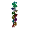
|
|---|---|
| 1 |
|
-要素
| #1: タンパク質 | 分子量: 41901.668 Da / 分子数: 12 / 由来タイプ: 天然 / 由来: (天然) #2: 化合物 | ChemComp-ADP /   分子量: 427.201 Da / 分子数: 12 / 由来タイプ: 合成 / 式: C10H15N5O10P2 / コメント: ADP, エネルギー貯蔵分子*YM 分子量: 427.201 Da / 分子数: 12 / 由来タイプ: 合成 / 式: C10H15N5O10P2 / コメント: ADP, エネルギー貯蔵分子*YM#3: 化合物 | ChemComp-PO4 /   分子量: 94.971 Da / 分子数: 36 / 由来タイプ: 合成 / 式: PO4 分子量: 94.971 Da / 分子数: 36 / 由来タイプ: 合成 / 式: PO4#4: 化合物 | ChemComp-MG /   分子量: 24.305 Da / 分子数: 72 / 由来タイプ: 合成 / 式: Mg 分子量: 24.305 Da / 分子数: 72 / 由来タイプ: 合成 / 式: Mg |
|---|
-実験情報
-実験
| 実験 | 手法: 電子顕微鏡法 |
|---|---|
| EM実験 | 試料の集合状態: FILAMENT / 3次元再構成法: 単粒子再構成法 |
- 試料調製
試料調製
| 構成要素 | 名称: actin filament in the presence of phosphate / タイプ: COMPLEX 詳細: 50 mM NaCl, 5 mM MgCl2, 0.025 mM ATP, 10 mM sodium phosphate (pH 7.4), 0.05 %NaN3, 0.7 mM DTT |
|---|---|
| 緩衝液 | 名称: phosphate buffer / pH: 7.4 / 詳細: phosphate buffer |
| 試料 | 包埋: NO / シャドウイング: NO / 染色: NO / 凍結: YES |
| 急速凍結 | 凍結剤: ETHANE / Temp: 77 K / 湿度: 90 % |
- 電子顕微鏡撮影
| 顕微鏡 | モデル: HITACHI EF2000 |
|---|---|
| 電子銃 | 電子線源:  FIELD EMISSION GUN / 加速電圧: 200 kV / 照射モード: SPOT SCAN FIELD EMISSION GUN / 加速電圧: 200 kV / 照射モード: SPOT SCAN |
| 電子レンズ | モード: BRIGHT FIELD / 倍率(公称値): 100000 X / 最大 デフォーカス(公称値): 1500 nm / 最小 デフォーカス(公称値): 1000 nm |
| 試料ホルダ | 試料ホルダーモデル: GATAN LIQUID NITROGEN 資料ホルダタイプ: Side entry liquid nitrogen-cooled cryo specimen holder 温度: 77 K / 最高温度: 77 K / 最低温度: 77 K / 傾斜角・最大: 0 ° / 傾斜角・最小: 0 ° |
| 撮影 | 電子線照射量: 15 e/Å2 / フィルム・検出器のモデル: GENERIC CCD 詳細: CCD camera with an epitaxially-grown CsI scintillator |
| 電子光学装置 | エネルギーフィルター名称: In-column Omega Filter |
- 解析
解析
| EMソフトウェア |
| ||||||||||||||||
|---|---|---|---|---|---|---|---|---|---|---|---|---|---|---|---|---|---|
| CTF補正 | 詳細: FSC at 0.143 cut-off | ||||||||||||||||
| 3次元再構成 | 手法: single particle / 解像度: 6 Å / 粒子像の数: 8000 / ピクセルサイズ(公称値): 2.275 Å / ピクセルサイズ(実測値): 2.275 Å / 対称性のタイプ: HELICAL | ||||||||||||||||
| 原子モデル構築 | プロトコル: FLEXIBLE FIT / 空間: REAL / Target criteria: pdbRhofit (Yasunaga & Wakabayashi, 1996) 詳細: METHOD--local refinement REFINEMENT PROTOCOL--real-space refinement | ||||||||||||||||
| 精密化ステップ | サイクル: LAST
|
