 ムービー
ムービー コントローラー


+ データを開く

- 基本情報
基本情報
| 登録情報 | データベース: PDB / ID: 2fvo | ||||||
|---|---|---|---|---|---|---|---|
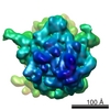
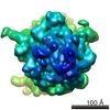
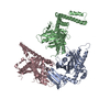
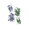
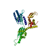
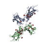
| タイトル | Docking of the modified RF1 X-ray structure into the Low Resolution Cryo-EM map of E.coli 70S Ribosome bound with RF1 | ||||||
 要素 要素 | Peptide chain release factor 1 | ||||||
 キーワード キーワード | TRANSLATION / RF1 ribosome cryo-EM | ||||||
| 機能・相同性 |  機能・相同性情報 機能・相同性情報 | ||||||
| 生物種 |   Thermotoga maritima (バクテリア) Thermotoga maritima (バクテリア) | ||||||
| 手法 | 電子顕微鏡法 / 単粒子再構成法 / クライオ電子顕微鏡法 / 解像度: 12.8 Å | ||||||
 データ登録者 データ登録者 | Rawat, U. / Gao, H. / Zavialov, A. / Gursky, R. / Ehrenberg, M. / Frank, J. | ||||||
 引用 引用 | ジャーナル: J Mol Biol / 年: 2006 タイトル: Interactions of the release factor RF1 with the ribosome as revealed by cryo-EM. 著者: Urmila Rawat / Haixiao Gao / Andrey Zavialov / Richard Gursky / Måns Ehrenberg / Joachim Frank /  要旨: In eubacteria, termination of translation is signaled by any one of the stop codons UAA, UAG, and UGA moving into the ribosomal A site. Two release factors, RF1 and RF2, recognize and bind to the ...In eubacteria, termination of translation is signaled by any one of the stop codons UAA, UAG, and UGA moving into the ribosomal A site. Two release factors, RF1 and RF2, recognize and bind to the stop codons with different affinities and trigger the hydrolysis of the ester bond that links the polypeptide with the P-site tRNA. Cryo-electron microscopy (cryo-EM) results obtained in this study show that ribosome-bound RF1 is in an open conformation, unlike the closed conformation observed in the crystal structure of the free factor, allowing its simultaneous access to both the decoding center and the peptidyl-transferase center. These results are similar to those obtained for RF2, but there is an important difference in how the factors bind to protein L11, which forms part of the GTPase-associated center of the large ribosomal subunit. The difference in the binding position, C-terminal domain for RF2 versus N-terminal domain for RF1, explains a body of L11 mutation studies that revealed differential effects on the activity of the two factors. Very recent data obtained with small-angle X-ray scattering now reveal that the solution structure of RF1 is open, as here seen on the ribosome by cryo-EM, and not closed, as seen in the crystal. #1: ジャーナル: J Mol Biol / 年: 2004タイトル: Structural analyses of peptide release factor 1 from Thermotoga maritima reveal domain flexibility required for its interaction with the ribosome. 著者: Dong Hae Shin / Jeroen Brandsen / Jaru Jancarik / Hisao Yokota / Rosalind Kim / Sung-Hou Kim / 要旨: We have determined the crystal structure of peptide chain release factor 1 (RF1) from Thermotoga maritima (gi 4981173) at 2.65 Angstrom resolution by selenomethionine single-wavelength anomalous ...We have determined the crystal structure of peptide chain release factor 1 (RF1) from Thermotoga maritima (gi 4981173) at 2.65 Angstrom resolution by selenomethionine single-wavelength anomalous dispersion (SAD) techniques. RF1 is a protein that recognizes stop codons and promotes the release of a nascent polypeptide from tRNA on the ribosome. Selenomethionine-labeled RF1 crystallized in space group P2(1) with three monomers per asymmetric unit. It has approximate dimensions of 75 Angstrom x 70 Angstrom x 45 Angstrom and is composed of four domains. The overall fold of each RF1 domain shows almost the same topology with Escherichia coli RF2, except that the RF1 N-terminal domain is shorter and the C-terminal domain is longer than that of RF2. The N-terminal domain of RF1 indicates a rigid-body movement relative to that of RF2 with an angle of approximately 90 degrees. Including these features, RF1 has a tripeptide anticodon PVT motif instead of the SPF motif of RF2, which confers the specificity towards the stop codons. The analyses of three molecules in the asymmetric unit and comparison with RF2 revealed the presence of dynamic movement of domains I and III, which are anchored to the central domain by hinge loops. The crystal structure of RF1 elucidates the intrinsic property of this family of having large domain movements for proper function with the ribosome. | ||||||
| 履歴 |
|
- 構造の表示
構造の表示
| ムービー |
ムービービューア |
|---|---|
| 構造ビューア | 分子: MolmilJmol/JSmol |
- ダウンロードとリンク
-ダウンロード
| PDBx/mmCIF形式 | 2fvo.cif.gz | 22.8 KB | 表示 | PDBx/mmCIF形式 |
|---|---|---|---|---|
| PDB形式 | pdb2fvo.ent.gz | 10.4 KB | 表示 | PDB形式 |
| PDBx/mmJSON形式 | 2fvo.json.gz | ツリー表示 | PDBx/mmJSON形式 | |
| その他 |  その他のダウンロード その他のダウンロード |
-検証レポート
| アーカイブディレクトリ | https://data.pdbj.org/pub/pdb/validation_reports/fv/2fvoftp://data.pdbj.org/pub/pdb/validation_reports/fv/2fvo | HTTPS FTP |
|---|
-関連構造データ


-リンク
 PDBj
PDBj

- 集合体
集合体
| 登録構造単位 | 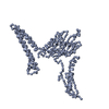
|
|---|---|
| 1 |
|
-要素
| #1: タンパク質 | 分子量: 38843.820 Da / 分子数: 1 / 由来タイプ: 組換発現 由来: (組換発現) Thermotoga maritima (バクテリア)遺伝子: prfA / 発現宿主: |
|---|
-実験情報
-実験
| 実験 | 手法: 電子顕微鏡法 |
|---|---|
| EM実験 | 試料の集合状態: PARTICLE / 3次元再構成法: 単粒子再構成法 |
- 試料調製
試料調製
| 構成要素 | 名称: E.COLI 70S RIBOSOME - RF2 complex / タイプ: RIBOSOME |
|---|---|
| 緩衝液 | 名称: Polymix buffer / pH: 7.5 / 詳細: Polymix buffer |
| 試料 | 濃度: 32 mg/ml / 包埋: NO / シャドウイング: NO / 染色: NO / 凍結: YES |
| 試料支持 | 詳細: quanti-foil grids coated with a thin carbon layer |
| 急速凍結 | 装置: HOMEMADE PLUNGER / 凍結剤: ETHANE / 詳細: RAPID-FREEZING IN LIQUID Ethane |
- 電子顕微鏡撮影
| 実験機器 |  モデル: Tecnai F20 / 画像提供: FEI Company |
|---|---|
| 顕微鏡 | モデル: FEI TECNAI F20 / 日付: 2002年12月1日 |
| 電子銃 | 電子線源:  FIELD EMISSION GUN / 加速電圧: 200 kV / 照射モード: FLOOD BEAM FIELD EMISSION GUN / 加速電圧: 200 kV / 照射モード: FLOOD BEAM |
| 電子レンズ | モード: BRIGHT FIELD / 倍率(公称値): 50000 X / 倍率(補正後): 49696 X / 最大 デフォーカス(公称値): 4000 nm / 最小 デフォーカス(公称値): 2000 nm / Cs: 2 mm |
| 試料ホルダ | 温度: 93 K / 傾斜角・最大: 0 ° / 傾斜角・最小: 0 ° |
| 撮影 | 電子線照射量: 20 e/Å2 / フィルム・検出器のモデル: KODAK SO-163 FILM |
- 解析
解析
| EMソフトウェア |
| ||||||||||||
|---|---|---|---|---|---|---|---|---|---|---|---|---|---|
| CTF補正 | 詳細: CTF CORRECTION OF 3D-MAPS | ||||||||||||
| 対称性 | 点対称性: C1 (非対称) | ||||||||||||
| 3次元再構成 | 手法: Single particle / 解像度: 12.8 Å / 解像度の算出法: FSC 0.5 CUT-OFF / 粒子像の数: 24622 / ピクセルサイズ(実測値): 2.82 Å / 倍率補正: TMV 詳細: Coordinates contain CA atoms only. ALl the image processing was done in SPIDER. 0.5 cutoff of FSC 対称性のタイプ: POINT | ||||||||||||
| 原子モデル構築 | プロトコル: OTHER / 空間: REAL / 詳細: METHOD--Manual | ||||||||||||
| 原子モデル構築 | PDB-ID: 1RQ0 Accession code: 1RQ0 / Source name: PDB / タイプ: experimental model | ||||||||||||
| 精密化 | 最高解像度: 12.8 Å / 詳細: Coordinates contain CA atoms only | ||||||||||||
| 精密化ステップ | サイクル: LAST / 最高解像度: 12.8 Å
|
