 Movie
Movie Controller

[English] 日本語
 Yorodumi
- PDB-1z7z: Cryo-em structure of human coxsackievirus A21 complexed with five... -
+ Open data

- Basic information
| Entry | Database: PDB / ID: 1z7z | |||||||||
|---|---|---|---|---|---|---|---|---|---|---|
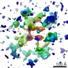
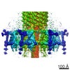
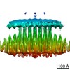
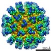
| Title | Cryo-em structure of human coxsackievirus A21 complexed with five domain icam-1kilifi | |||||||||
 Components Components |
| |||||||||
 Keywords Keywords | Virus/Receptor / ICAM-1 / Kilifi / CD54 / Human Coxsackievirus A21 / virus-receptor complex / Icosahedral virus | |||||||||
| Function / homology |  Function and homology information Function and homology informationregulation of leukocyte mediated cytotoxicity / T cell extravasation / positive regulation of cellular extravasation / leukocyte adhesion to vascular endothelial cell / regulation of ruffle assembly / membrane to membrane docking / T cell antigen processing and presentation / cell-cell adhesion mediated by integrin / T cell activation via T cell receptor contact with antigen bound to MHC molecule on antigen presenting cell / adhesion of symbiont to host ...regulation of leukocyte mediated cytotoxicity / T cell extravasation / positive regulation of cellular extravasation / leukocyte adhesion to vascular endothelial cell / regulation of ruffle assembly / membrane to membrane docking / T cell antigen processing and presentation / cell-cell adhesion mediated by integrin / T cell activation via T cell receptor contact with antigen bound to MHC molecule on antigen presenting cell / adhesion of symbiont to host / establishment of endothelial barrier / heterophilic cell-cell adhesion / leukocyte migration / leukocyte cell-cell adhesion / Interleukin-10 signaling / symbiont-mediated suppression of host cytoplasmic pattern recognition receptor signaling pathway via inhibition of RIG-I activity / immunological synapse / Integrin cell surface interactions / negative regulation of endothelial cell apoptotic process / negative regulation of extrinsic apoptotic signaling pathway via death domain receptors / cellular response to leukemia inhibitory factor / picornain 2A / symbiont-mediated suppression of host mRNA export from nucleus / symbiont genome entry into host cell via pore formation in plasma membrane / picornain 3C / cellular response to glucose stimulus / T=pseudo3 icosahedral viral capsid / host cell cytoplasmic vesicle membrane / integrin binding / cellular response to amyloid-beta / Interferon gamma signaling / Immunoregulatory interactions between a Lymphoid and a non-Lymphoid cell / ribonucleoside triphosphate phosphatase activity / transmembrane signaling receptor activity / nucleoside-triphosphate phosphatase / virus receptor activity / extracellular matrix / channel activity / signaling receptor activity / Interleukin-4 and Interleukin-13 signaling / monoatomic ion transmembrane transport / receptor-mediated virion attachment to host cell / positive regulation of ERK1 and ERK2 cascade / DNA replication / cell adhesion / RNA helicase activity / endocytosis involved in viral entry into host cell / membrane raft / receptor ligand activity / external side of plasma membrane / symbiont-mediated activation of host autophagy / RNA-directed RNA polymerase / cysteine-type endopeptidase activity / focal adhesion / viral RNA genome replication / RNA-directed RNA polymerase activity / DNA-templated transcription / virion attachment to host cell / host cell nucleus / structural molecule activity / cell surface / proteolysis / : / RNA binding / extracellular exosome / zinc ion binding / ATP binding / membrane / plasma membrane Similarity search - Function | |||||||||
| Biological species |  Human coxsackievirus A21 Human coxsackievirus A21 Homo sapiens (human) Homo sapiens (human) | |||||||||
| Method | ELECTRON MICROSCOPY / single particle reconstruction / cryo EM / Resolution: 8 Å | |||||||||
 Authors Authors | Xiao, C. / Bator-Kelly, C.M. / Rieder, E. / Chipman, P.R. / Craig, A. / Kuhn, R.J. / Wimmer, E. / Rossmann, M.G. | |||||||||
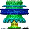
 Citation Citation | Journal: Structure / Year: 2005 Title: The crystal structure of coxsackievirus A21 and its interaction with ICAM-1. Authors: Chuan Xiao / Carol M Bator-Kelly / Elizabeth Rieder / Paul R Chipman / Alister Craig / Richard J Kuhn / Eckard Wimmer / Michael G Rossmann /  Abstract: CVA21 and polioviruses both belong to the Enterovirus genus in the family of Picornaviridae, whereas rhinoviruses form a distinct picornavirus genus. Nevertheless, CVA21 and the major group of human ...CVA21 and polioviruses both belong to the Enterovirus genus in the family of Picornaviridae, whereas rhinoviruses form a distinct picornavirus genus. Nevertheless, CVA21 and the major group of human rhinoviruses recognize intercellular adhesion molecule-1 (ICAM-1) as their cellular receptor, whereas polioviruses use poliovirus receptor. The crystal structure of CVA21 has been determined to 3.2 A resolution. Its structure has greater similarity to poliovirus structures than to other known picornavirus structures. Cryo-electron microscopy (cryo-EM) was used to determine an 8.0 A resolution structure of CVA21 complexed with an ICAM-1 variant, ICAM-1(Kilifi). The cryo-EM map was fitted with the crystal structures of ICAM-1 and CVA21. Significant differences in the structure of CVA21 with respect to the poliovirus structures account for the inability of ICAM-1 to bind polioviruses. The interface between CVA21 and ICAM-1 has shape and electrostatic complementarity with many residues being conserved among those CVAs that bind ICAM-1. | |||||||||
| History |
|
- Structure visualization
| Movie |
Movie viewer |
|---|---|
| Structure viewer | Molecule: MolmilJmol/JSmol |
- Downloads & links
-Download
| PDBx/mmCIF format | 1z7z.cif.gz | 212.7 KB | Display | PDBx/mmCIF format |
|---|---|---|---|---|
| PDB format | pdb1z7z.ent.gz | 159.5 KB | Display | PDB format |
| PDBx/mmJSON format | 1z7z.json.gz | Tree view | PDBx/mmJSON format | |
| Others |  Other downloads Other downloads |
-Validation report
| Arichive directory | https://data.pdbj.org/pub/pdb/validation_reports/z7/1z7zftp://data.pdbj.org/pub/pdb/validation_reports/z7/1z7z | HTTPS FTP |
|---|
-Related structure data
| Related structure data | 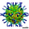 1114MC 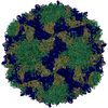 1z7sC C: citing same article ( M: map data used to model this data |
|---|---|
| Similar structure data |
-Links
 PDBj
PDBj


- Assembly
| Deposited unit | 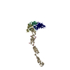
|
|---|---|
| 1 | x 60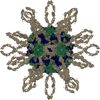
|
| 2 |
|
| 3 | x 5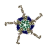
|
| 4 | x 6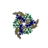
|
| 5 | 
|
| Symmetry | Point symmetry: (Hermann–Mauguin notation: 532 / Schoenflies symbol: I (icosahedral)) |
-Components
-Human coxsackievirus ... , 5 types, 5 molecules 12345
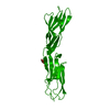
| #1: Protein | Mass: 31946.727 Da / Num. of mol.: 1 / Fragment: Viral Protein 1 residues 1073-1286 / Source method: isolated from a natural source / Details: THE NATURE HOST OF THIS VIRUS IS HUMAN / Source: (natural) Human coxsackievirus A21 / Genus: Enterovirus / Cell line: Hela / Species: Human enterovirus C / Strain: KUYKENDALL / References: GenBank: 33304569, UniProt: Q7T7N6*PLUS |
|---|---|
| #2: Protein | Mass: 29918.537 Da / Num. of mol.: 1 / Fragment: Viral Protein 2 residues 2010-2272 / Source method: isolated from a natural source / Details: THE NATURE HOST OF THIS VIRUS IS HUMAN / Source: (natural) Human coxsackievirus A21 / Genus: Enterovirus / Cell line: Hela / Species: Human enterovirus C / Strain: KUYKENDALL / References: GenBank: 33304569, UniProt: Q7T7N6*PLUS |
| #3: Protein | Mass: 25898.895 Da / Num. of mol.: 1 / Fragment: Viral Protein 3 residues 3043-3234 / Source method: isolated from a natural source / Details: THE NATURE HOST OF THIS VIRUS IS HUMAN / Source: (natural) Human coxsackievirus A21 / Genus: Enterovirus / Cell line: Hela / Species: Human enterovirus C / Strain: KUYKENDALL / References: GenBank: 33304569, UniProt: Q7T7N6*PLUS |
| #4: Protein/peptide | Mass: 1335.565 Da / Num. of mol.: 1 / Fragment: Viral Protein 1 residues 1287-1298 / Source method: isolated from a natural source / Details: THE NATURE HOST OF THIS VIRUS IS HUMAN / Source: (natural) Human coxsackievirus A21 / Genus: Enterovirus / Cell line: Hela / Species: Human enterovirus C / Strain: KUYKENDALL / References: GenBank: 33304569, UniProt: P22055*PLUS |
| #5: Protein/peptide | Mass: 687.809 Da / Num. of mol.: 1 / Fragment: Viral Protein 3 residues 3035-3239 / Source method: isolated from a natural source / Details: THE NATURE HOST OF THIS VIRUS IS HUMAN / Source: (natural) Human coxsackievirus A21 / Genus: Enterovirus / Cell line: Hela / Species: Human enterovirus C / Strain: KUYKENDALL / References: GenBank: 33304569 |
-Protein / Sugars , 2 types, 9 molecules I
| #6: Protein | Mass: 49104.473 Da / Num. of mol.: 1 / Fragment: ICAM-1 EXTRACELLULAR DOMAIN 1-5 / Mutation: K29M / Source method: isolated from a natural source / Source: (natural) Homo sapiens (human) / Cell line: COS7 / References: UniProt: P05362 |
|---|---|
| #7: Sugar | ChemComp-NAG /  Type: D-saccharide, beta linking / Mass: 221.208 Da / Num. of mol.: 8 Type: D-saccharide, beta linking / Mass: 221.208 Da / Num. of mol.: 8Source method: isolated from a genetically manipulated source Formula: C8H15NO6 |
-Details
| Has protein modification | Y |
|---|
-Experimental details
-Experiment
| Experiment | Method: ELECTRON MICROSCOPY |
|---|---|
| EM experiment | Aggregation state: PARTICLE / 3D reconstruction method: single particle reconstruction |
- Sample preparation
| Component |
| ||||||||||||||||||||||||||||||||||||||||
|---|---|---|---|---|---|---|---|---|---|---|---|---|---|---|---|---|---|---|---|---|---|---|---|---|---|---|---|---|---|---|---|---|---|---|---|---|---|---|---|---|---|
| Details of virus | Host category: MAMMALIAN / Type: VIRION | ||||||||||||||||||||||||||||||||||||||||
| Natural host | Organism: Homo sapiens / Strain: Hela | ||||||||||||||||||||||||||||||||||||||||
| Buffer solution | Name: TRIS / pH: 7.2 / Details: TRIS | ||||||||||||||||||||||||||||||||||||||||
| Specimen | Conc.: 5 mg/ml / Embedding applied: NO / Shadowing applied: NO / Staining applied: NO / Vitrification applied: YES | ||||||||||||||||||||||||||||||||||||||||
| Specimen support | Details: HOLEY CARBON | ||||||||||||||||||||||||||||||||||||||||
| Vitrification | Instrument: REICHERT-JUNG PLUNGER / Cryogen name: ETHANE / Details: PLUNGED INTO ETHANE AT LIQUID NITROGEN TEMPERATURE |
- Electron microscopy imaging
| Microscopy | Model: FEI/PHILIPS CM300FEG/T / Date: Mar 15, 2001 Details: SAMPLES WERE MAINTAINED AT LIQUID NITROGEN TEMPERATURES IN THE ELECTRON MICROSCOPE. |
|---|---|
| Electron gun | Electron source:  FIELD EMISSION GUN / Accelerating voltage: 300 kV / Illumination mode: OTHER FIELD EMISSION GUN / Accelerating voltage: 300 kV / Illumination mode: OTHER |
| Electron lens | Mode: BRIGHT FIELD / Nominal magnification: 45000 X / Calibrated magnification: 47000 X / Nominal defocus max: 3000 nm / Nominal defocus min: 700 nm / Cs: 2 mm |
| Specimen holder | Temperature: 100 K / Tilt angle max: 0 ° / Tilt angle min: 0 ° |
| Image recording | Electron dose: 26 e/Å2 / Film or detector model: KODAK SO-163 FILM |
| Image scans | Num. digital images: 33 |
| Radiation | Protocol: SINGLE WAVELENGTH / Monochromatic (M) / Laue (L): M / Scattering type: x-ray |
| Radiation wavelength | Relative weight: 1 |
- Processing
| EM software |
| |||||||||||||||||||||
|---|---|---|---|---|---|---|---|---|---|---|---|---|---|---|---|---|---|---|---|---|---|---|
| CTF correction | Details: REVERSED CTF WITH WEINER FACTOR FOR EACH PARTICLE | |||||||||||||||||||||
| Symmetry | Point symmetry: I (icosahedral) | |||||||||||||||||||||
| 3D reconstruction | Method: POLAR FOURIER TRANSFORM METHOD AND FOURIER-BESSEL RECONSTRUCTION Resolution: 8 Å / Num. of particles: 4704 / Nominal pixel size: 2.98 Å / Actual pixel size: 2.85 Å Magnification calibration: THE PIXEL SIZE OF THE CRYO-EM MAP WAS CALIBRATED AGAINST THE ATOMIC MODEL OF THE VIRUS CAPSID. DENSITIES WERE COMPARED BY CROSS- CORRELATION WITHIN A SPHERICAL SHELL OF ...Magnification calibration: THE PIXEL SIZE OF THE CRYO-EM MAP WAS CALIBRATED AGAINST THE ATOMIC MODEL OF THE VIRUS CAPSID. DENSITIES WERE COMPARED BY CROSS- CORRELATION WITHIN A SPHERICAL SHELL OF INTERNAL RADIUS 104 ANGSTROMS AND EXTERNAL RADIUS 177 ANGSTROMS. Details: A B-FACTOR OF 589.46 WAS SET FOR ALL THE ATOMS BASED ON THE EQUATION OF -4*LN(0.1*RESOLUTION2) TO MIMIC A 8 ANGSTROM RESOLUTION CRYOEM STRUCTURE. OCCUPANCY OF 1.0 WAS SET FOR ALL ATOMS OF ...Details: A B-FACTOR OF 589.46 WAS SET FOR ALL THE ATOMS BASED ON THE EQUATION OF -4*LN(0.1*RESOLUTION2) TO MIMIC A 8 ANGSTROM RESOLUTION CRYOEM STRUCTURE. OCCUPANCY OF 1.0 WAS SET FOR ALL ATOMS OF THE VIRAL CAPSID. OCCUPANCY OF 0.80, 0.56, 0.24, 0.16, AND 0.08 WAS SET FOR ATOMS OF ICAM-1 DOMAIN 1, 2, 3, 4, AND 5, RESPECTIVELY, BASED ON THE FITTING RESULTS. SEE DETAILS IN THE CITATION. Symmetry type: POINT | |||||||||||||||||||||
| Atomic model building | Protocol: RIGID BODY FIT / Space: REAL Target criteria: BEST SUMF VALUE FIT USING THE PROGRAM EMFIT Details: REFINEMENT PROTOCOL--RIGID BODY | |||||||||||||||||||||
| Atomic model building |
| |||||||||||||||||||||
| Refinement | Highest resolution: 8 Å | |||||||||||||||||||||
| Refinement step | Cycle: LAST / Highest resolution: 8 Å
|
