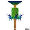
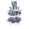
Journal: Cell / Year: 2004 Title: Three-dimensional rearrangement of proteins in the tail of bacteriophage T4 on infection of its host. Authors: Petr G Leiman / Paul R Chipman / Victor A Kostyuchenko / Vadim V Mesyanzhinov / Michael G Rossmann / Abstract: The contractile tail of bacteriophage T4 undergoes major structural transitions when the virus attaches to the host cell surface. The baseplate at the distal end of the tail changes from a hexagonal ...The contractile tail of bacteriophage T4 undergoes major structural transitions when the virus attaches to the host cell surface. The baseplate at the distal end of the tail changes from a hexagonal to a star shape. This causes the sheath around the tail tube to contract and the tail tube to protrude from the baseplate and pierce the outer cell membrane and the cell wall before reaching the inner cell membrane for subsequent viral DNA injection. Analogously, the T4 tail can be contracted by treatment with 3 M urea. The structure of the T4 contracted tail, including the head-tail joining region, has been determined by cryo-electron microscopy to 17 A resolution. This 1200 A-long, 20 MDa structure has been interpreted in terms of multiple copies of its approximately 20 component proteins. A comparison with the metastable hexagonal baseplate of the mature virus shows that the baseplate proteins move as rigid bodies relative to each other during the structural change.
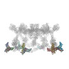
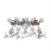
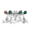
A: Baseplate structural protein Gp8 B: Baseplate structural protein Gp8 C: Baseplate structural protein Gp9 D: Baseplate structural protein Gp9 E: Baseplate structural protein Gp9 F: Baseplate structural protein Gp11 G: Baseplate structural protein Gp11 H: Baseplate structural protein Gp11
A: Baseplate structural protein Gp8 B: Baseplate structural protein Gp8 C: Baseplate structural protein Gp9 D: Baseplate structural protein Gp9 E: Baseplate structural protein Gp9 F: Baseplate structural protein Gp11 G: Baseplate structural protein Gp11 H: Baseplate structural protein Gp11
Electron dose: 20 e/Å2 / Film or detector model: KODAK SO-163 FILM
-
Processing
EM software
ID
Name
Version
Category
1
Situs
modelfitting
2
SPIDER
2
3Dreconstruction
CTF correction
Details: CTF correction of each particle
Symmetry
Point symmetry: C6 (6 fold cyclic)
3D reconstruction
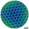
Method: Back projection / Resolution: 16 Å / Num. of particles: 1965 / Nominal pixel size: 4.10442 Å / Actual pixel size: 3.93285 Å / Magnification calibration: Catalase crystals diffraction / Symmetry type: POINT
Atomic model building
Protocol: OTHER / Space: REAL / Details: METHOD--Laplacian filtered real space
In the structure databanks used in Yorodumi, some data are registered as the other names, "COVID-19 virus" and "2019-nCoV". Here are the details of the virus and the list of structure data.
Jan 31, 2019. EMDB accession codes are about to change! (news from PDBe EMDB page)
EMDB accession codes are about to change! (news from PDBe EMDB page)
The allocation of 4 digits for EMDB accession codes will soon come to an end. Whilst these codes will remain in use, new EMDB accession codes will include an additional digit and will expand incrementally as the available range of codes is exhausted. The current 4-digit format prefixed with “EMD-” (i.e. EMD-XXXX) will advance to a 5-digit format (i.e. EMD-XXXXX), and so on. It is currently estimated that the 4-digit codes will be depleted around Spring 2019, at which point the 5-digit format will come into force.
The EM Navigator/Yorodumi systems omit the EMD- prefix.
Related info.:Q: What is EMD? / ID/Accession-code notation in Yorodumi/EM Navigator
Yorodumi is a browser for structure data from EMDB, PDB, SASBDB, etc.
This page is also the successor to EM Navigator detail page, and also detail information page/front-end page for Omokage search.
The word "yorodu" (or yorozu) is an old Japanese word meaning "ten thousand". "mi" (miru) is to see.
Related info.:EMDB / PDB / SASBDB / Comparison of 3 databanks / Yorodumi Search / Aug 31, 2016. New EM Navigator & Yorodumi / Yorodumi Papers / Jmol/JSmol / Function and homology information / Changes in new EM Navigator and Yorodumi
 Movie
Movie Controller

 Yorodumi
 Open data
 Basic information
 Components
 Keywords
 Function and homology information
 Enterobacteria phage T4 (virus)
 Authors
 Citation
 Structure visualization
 Downloads & links
 Other downloads


 PDBj
PDBj Assembly
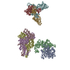


 Sample preparation
 Electron microscopy imaging
 FIELD EMISSION GUN / Accelerating voltage: 300 kV / Illumination mode: SPOT SCAN
 Processing