Movie
Movie Controller

[English] 日本語
 Yorodumi
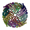
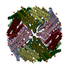
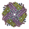
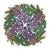
- EMDB-9910: Cryo-EM structure of Apo-bacterioferritin from Streptomyces coelicolor -
+ Open data

- Basic information
| Entry | Database: EMDB / ID: EMD-9910 | |||||||||
|---|---|---|---|---|---|---|---|---|---|---|
| Title | Cryo-EM structure of Apo-bacterioferritin from Streptomyces coelicolor | |||||||||
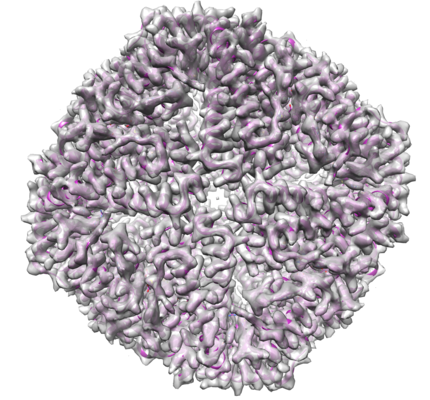
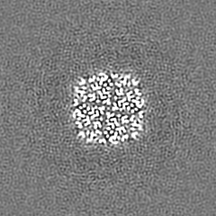
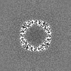
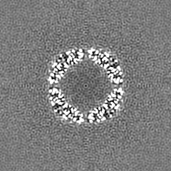
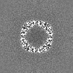
 Map data Map data | The cryo-EM map of Apo-bacterioferritin from Streptomyces coelicolor | |||||||||
 Sample Sample |
| |||||||||
 Keywords Keywords | Ferritin / Nanocage / Bacterioferritin / METAL TRANSPORT | |||||||||
| Function / homology |  Function and homology information Function and homology informationferroxidase / ferroxidase activity / ferric iron binding / iron ion transport / intracellular iron ion homeostasis / iron ion binding / heme binding / cytosol Similarity search - Function | |||||||||
| Biological species |  Streptomyces coelicolor (bacteria) Streptomyces coelicolor (bacteria) | |||||||||
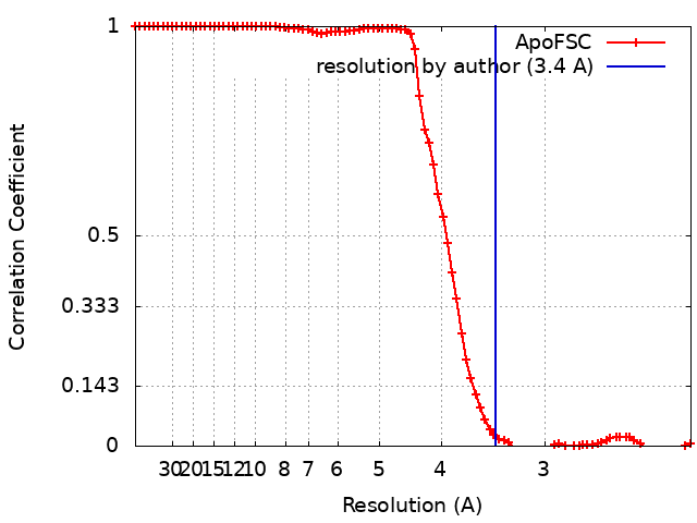
| Method | single particle reconstruction / cryo EM / Resolution: 3.4 Å | |||||||||
 Authors Authors | Jobichen C / Sivaraman J | |||||||||
 Citation Citation | Journal: To Be Published Title: Cryo-EM structure of Apo-bacterioferritin from Streptomyces coelicolor. Authors: Jobichen C / Sivaraman J | |||||||||
| History |
|
- Structure visualization
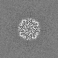
| Movie |
Movie viewer |
|---|---|
| Structure viewer | EM map: SurfViewMolmilJmol/JSmol |
| Supplemental images |
- Downloads & links
-EMDB archive
| Map data | emd_9910.map.gz | 49 MB | EMDB map data format | |
|---|---|---|---|---|
| Header (meta data) | emd-9910-v30.xmlemd-9910.xml | 13.3 KB 13.3 KB | Display Display | EMDB header |
| FSC (resolution estimation) | emd_9910_fsc.xml | 7.8 KB | Display | FSC data file |
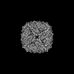
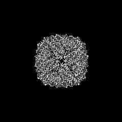
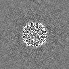
| Images |  emd_9910.png emd_9910.png | 273.2 KB | ||
| Filedesc metadata | emd-9910.cif.gz | 5.6 KB | ||
| Archive directory |  http://ftp.pdbj.org/pub/emdb/structures/EMD-9910ftp://ftp.pdbj.org/pub/emdb/structures/EMD-9910 http://ftp.pdbj.org/pub/emdb/structures/EMD-9910ftp://ftp.pdbj.org/pub/emdb/structures/EMD-9910 | HTTPS FTP |
-Related structure data
| Related structure data |  6k3oMC  9913C 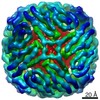 9915C  6k43C 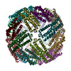 6k4mC M: atomic model generated by this map C: citing same article ( |
|---|---|
| Similar structure data |


-Links
| EMDB pages | EMDB (EBI/PDBe) / EMDataResource |
|---|---|
| Related items in Molecule of the Month |


-Map
| File | Download / File: emd_9910.map.gz / Format: CCP4 / Size: 52.7 MB / Type: IMAGE STORED AS FLOATING POINT NUMBER (4 BYTES) | ||||||||||||||||||||||||||||||||||||||||||||||||||||||||||||||||||||
|---|---|---|---|---|---|---|---|---|---|---|---|---|---|---|---|---|---|---|---|---|---|---|---|---|---|---|---|---|---|---|---|---|---|---|---|---|---|---|---|---|---|---|---|---|---|---|---|---|---|---|---|---|---|---|---|---|---|---|---|---|---|---|---|---|---|---|---|---|---|
| Annotation | The cryo-EM map of Apo-bacterioferritin from Streptomyces coelicolor | ||||||||||||||||||||||||||||||||||||||||||||||||||||||||||||||||||||
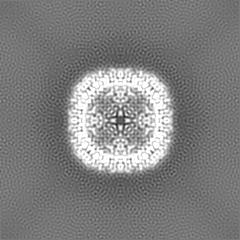
| Projections & slices | Image control
Images are generated by Spider. | ||||||||||||||||||||||||||||||||||||||||||||||||||||||||||||||||||||
| Voxel size | X=Y=Z: 1.11 Å | ||||||||||||||||||||||||||||||||||||||||||||||||||||||||||||||||||||
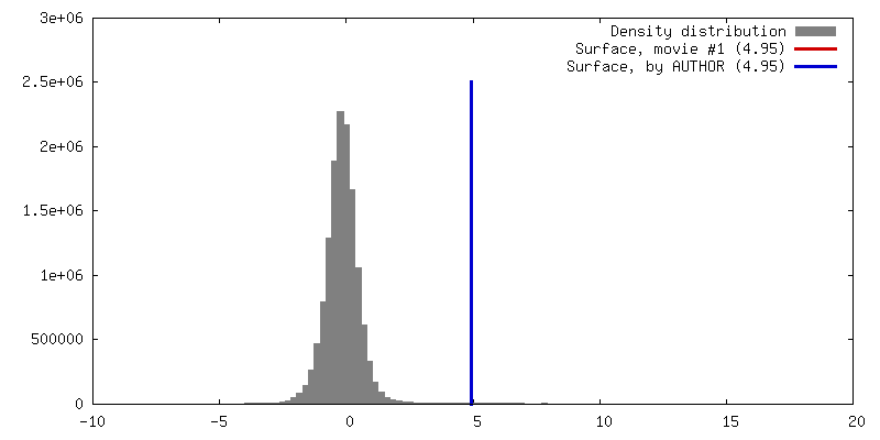
| Density |
| ||||||||||||||||||||||||||||||||||||||||||||||||||||||||||||||||||||
| Symmetry | Space group: 1 | ||||||||||||||||||||||||||||||||||||||||||||||||||||||||||||||||||||
| Details | EMDB XML:
CCP4 map header:
| ||||||||||||||||||||||||||||||||||||||||||||||||||||||||||||||||||||
 Z (Sec.)
 Y (Row.)
 X (Col.)


-Supplemental data
- Sample components
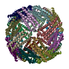
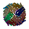
-Entire : Bacterioferritin in complex with HEME and IRON
| Entire | Name: Bacterioferritin in complex with HEME and IRON |
|---|---|
| Components |
|
-Supramolecule #1: Bacterioferritin in complex with HEME and IRON
| Supramolecule | Name: Bacterioferritin in complex with HEME and IRON / type: organelle_or_cellular_component / ID: 1 / Parent: 0 / Macromolecule list: #1 |
|---|---|
| Source (natural) | Organism: Streptomyces coelicolor (bacteria) |
| Molecular weight | Theoretical: 500 KDa |
-Macromolecule #1: Bacterioferritin
| Macromolecule | Name: Bacterioferritin / type: protein_or_peptide / ID: 1 / Number of copies: 24 / Enantiomer: LEVO / EC number: ferroxidase |
|---|---|
| Source (natural) | Organism: Streptomyces coelicolor (bacteria) |
| Molecular weight | Theoretical: 19.242629 KDa |
| Recombinant expression | Organism: |
| Sequence | String: MQGDPEVIEF LNEQLTAELT AINQYFLHAK LQDHKGWTKL AKYTRAESFD EMRHAEVLTD RILLLDGLPN YQRLFHVRVG QSVTEMFQA DREVELEAID RLRRGIEVMR AKHDITSANV FEAILADEEH HIDYLETQLD LIEKLGESLY LSTVIEQTQP D PSGPGSL UniProtKB: Bacterioferritin |
-Macromolecule #2: FE (II) ION
| Macromolecule | Name: FE (II) ION / type: ligand / ID: 2 / Number of copies: 24 / Formula: FE2 |
|---|---|
| Molecular weight | Theoretical: 55.845 Da |
-Macromolecule #3: PROTOPORPHYRIN IX CONTAINING FE
| Macromolecule | Name: PROTOPORPHYRIN IX CONTAINING FE / type: ligand / ID: 3 / Number of copies: 12 / Formula: HEM |
|---|---|
| Molecular weight | Theoretical: 616.487 Da |
| Chemical component information |  ChemComp-HEM: |
-Experimental details
-Structure determination
| Method | cryo EM |
|---|---|
 Processing Processing | single particle reconstruction |
| Aggregation state | particle |
-Sample preparation
| Concentration | 1.8 mg/mL |
|---|---|
| Buffer | pH: 7 |
| Grid | Model: Quantifoil, UltrAuFoil, R1.2/1.3 / Material: GOLD / Pretreatment - Type: GLOW DISCHARGE |
| Vitrification | Cryogen name: ETHANE |
| Details | This sample was monodisperse with 24mer nanocage assemblies. |
- Electron microscopy
| Microscope | FEI TITAN KRIOS |
|---|---|
| Image recording | Film or detector model: FEI FALCON II (4k x 4k) / Average electron dose: 1.6 e/Å2 |
| Electron beam | Acceleration voltage: 300 kV / Electron source:  FIELD EMISSION GUN FIELD EMISSION GUN |
| Electron optics | C2 aperture diameter: 2.7 µm / Illumination mode: OTHER / Imaging mode: BRIGHT FIELD |
| Experimental equipment |  Model: Titan Krios / Image courtesy: FEI Company |