Movie
Movie Controller

[English] 日本語
 Yorodumi
- PDB-6k43: Cryo-EM structure of Holo-bacterioferritin-form-I from Streptomyc... -
+ Open data

- Basic information
| Entry | Database: PDB / ID: 6k43 | ||||||
|---|---|---|---|---|---|---|---|
| Title | Cryo-EM structure of Holo-bacterioferritin-form-I from Streptomyces coelicolor | ||||||
 Components Components | Bacterioferritin | ||||||
 Keywords Keywords | METAL TRANSPORT / Ferritin / Nanocage / Bacterioferritin | ||||||
| Function / homology |  Function and homology information Function and homology informationferroxidase / ferroxidase activity / ferric iron binding / iron ion transport / intracellular iron ion homeostasis / iron ion binding / heme binding / cytosol Similarity search - Function | ||||||
| Biological species |  Streptomyces coelicolor (bacteria) Streptomyces coelicolor (bacteria) | ||||||
| Method | ELECTRON MICROSCOPY / single particle reconstruction / cryo EM / Resolution: 3.7 Å | ||||||
 Authors Authors | Jobichen, C. / Sivaraman, J. | ||||||
 Citation Citation | Journal: To Be Published Title: Cryo-EM structure of Apo-bacterioferritin from Streptomyces coelicolor. Authors: Jobichen, C. / Sivaraman, J. | ||||||
| History |
|
- Structure visualization
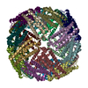
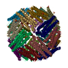
| Movie |
Movie viewer |
|---|---|
| Structure viewer | Molecule: MolmilJmol/JSmol |
- Downloads & links
-Download
| PDBx/mmCIF format | 6k43.cif.gz | 686.6 KB | Display | PDBx/mmCIF format |
|---|---|---|---|---|
| PDB format | pdb6k43.ent.gz | 580.4 KB | Display | PDB format |
| PDBx/mmJSON format | 6k43.json.gz | Tree view | PDBx/mmJSON format | |
| Others |  Other downloads Other downloads |
-Validation report
| Arichive directory | https://data.pdbj.org/pub/pdb/validation_reports/k4/6k43ftp://data.pdbj.org/pub/pdb/validation_reports/k4/6k43 | HTTPS FTP |
|---|
-Related structure data
| Related structure data |  9913MC 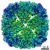 9910C 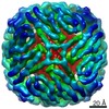 9915C 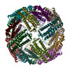 6k3oC 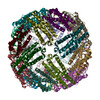 6k4mC C: citing same article ( M: map data used to model this data |
|---|---|
| Similar structure data |


-Links
 PDBj
PDBj


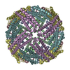
- Assembly
| Deposited unit | 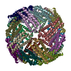
|
|---|---|
| 1 |
|
-Components
| #1: Protein | Mass: 19242.629 Da / Num. of mol.: 24 Source method: isolated from a genetically manipulated source Source: (gene. exp.) Streptomyces coelicolor (bacteria)Production host: Variant (production host): 'BL21-Gold(DE3)pLysS AG' / References: UniProt: Q9S2N0, ferroxidase #2: Chemical | ChemComp-FE2 /   Mass: 55.845 Da / Num. of mol.: 24 / Source method: obtained synthetically / Formula: Fe / Feature type: SUBJECT OF INVESTIGATION Mass: 55.845 Da / Num. of mol.: 24 / Source method: obtained synthetically / Formula: Fe / Feature type: SUBJECT OF INVESTIGATION#3: Chemical | ChemComp-HEM /   Mass: 616.487 Da / Num. of mol.: 12 / Source method: obtained synthetically / Formula: C34H32FeN4O4 / Feature type: SUBJECT OF INVESTIGATION Mass: 616.487 Da / Num. of mol.: 12 / Source method: obtained synthetically / Formula: C34H32FeN4O4 / Feature type: SUBJECT OF INVESTIGATION |
|---|
-Experimental details
-Experiment
| Experiment | Method: ELECTRON MICROSCOPY |
|---|---|
| EM experiment | Aggregation state: PARTICLE / 3D reconstruction method: single particle reconstruction |
- Sample preparation
| Component | Name: Bacterioferritin in complex with HEME with Fe3 mineral core Type: ORGANELLE OR CELLULAR COMPONENT / Entity ID: #1 / Source: RECOMBINANT |
|---|---|
| Molecular weight | Value: 0.5 MDa / Experimental value: YES |
| Source (natural) | Organism: Streptomyces coelicolor (bacteria) |
| Source (recombinant) | Organism: Strain: 'BL21-Gold(DE3)pLysS AG' |
| Buffer solution | pH: 7 |
| Specimen | Conc.: 1.8 mg/ml / Embedding applied: NO / Shadowing applied: NO / Staining applied: NO / Vitrification applied: YES Details: This sample was monodisperse with 24mer nanocage assemblies. |
| Vitrification | Cryogen name: ETHANE |
- Electron microscopy imaging
| Experimental equipment |  Model: Titan Krios / Image courtesy: FEI Company |
|---|---|
| Microscopy | Model: FEI TITAN KRIOS |
| Electron gun | Electron source:  FIELD EMISSION GUN / Accelerating voltage: 300 kV / Illumination mode: OTHER FIELD EMISSION GUN / Accelerating voltage: 300 kV / Illumination mode: OTHER |
| Electron lens | Mode: BRIGHT FIELD |
| Image recording | Electron dose: 1.6 e/Å2 / Film or detector model: FEI FALCON II (4k x 4k) |
- Processing
| Software | Name: PHENIX / Version: 1.15.2_3472: / Classification: refinement | ||||||||||||||||||||||||
|---|---|---|---|---|---|---|---|---|---|---|---|---|---|---|---|---|---|---|---|---|---|---|---|---|---|
| EM software |
| ||||||||||||||||||||||||
| CTF correction | Type: PHASE FLIPPING AND AMPLITUDE CORRECTION | ||||||||||||||||||||||||
| Symmetry | Point symmetry: O (octahedral) | ||||||||||||||||||||||||
| 3D reconstruction | Resolution: 3.7 Å / Resolution method: FSC 0.143 CUT-OFF / Num. of particles: 19485 / Symmetry type: POINT | ||||||||||||||||||||||||
| Atomic model building | Protocol: RIGID BODY FIT | ||||||||||||||||||||||||
| Atomic model building | PDB-ID: 5XX9 Accession code: 5XX9 / Source name: PDB / Type: experimental model | ||||||||||||||||||||||||
| Refine LS restraints |
|