ジャーナル: J Mol Biol / 年: 2019 タイトル: Cryo-EM Structures Reveal Relocalization of MetAP in the Presence of Other Protein Biogenesis Factors at the Ribosomal Tunnel Exit. 著者: Sayan Bhakta / Shirin Akbar / Jayati Sengupta / 要旨: During protein biosynthesis in bacteria, one of the earliest events that a nascent polypeptide chain goes through is the co-translational enzymatic processing. The event includes two enzymatic ...During protein biosynthesis in bacteria, one of the earliest events that a nascent polypeptide chain goes through is the co-translational enzymatic processing. The event includes two enzymatic pathways: deformylation of the N-terminal methionine by the enzyme peptide deformylase (PDF), followed by methionine excision catalyzed by methionine aminopeptidase (MetAP). During the enzymatic processing, the emerging nascent protein likely remains shielded by the ribosome-associated chaperone trigger factor. The ribosome tunnel exit serves as a stage for recruiting proteins involved in maturation processes of the nascent chain. Co-translational processing of nascent chains is a critical step for subsequent folding and functioning of mature proteins. Here, we present cryo-electron microscopy structures of Escherichia coli (E. coli) ribosome in complex with the nascent chain processing proteins. The structures reveal overlapping binding sites for PDF and MetAP when they bind individually at the tunnel exit site, where L22-L32 protein region provides primary anchoring sites for both proteins. In the absence of PDF, trigger factor can access ribosomal tunnel exit when MetAP occupies its primary binding site. Interestingly, however, in the presence of PDF, when MetAP's primary binding site is already engaged, MetAP has a remarkable ability to occupy an alternative binding site adjacent to PDF. Our study, thus, discloses an unexpected mechanism that MetAP adopts for context-specific ribosome association.
ダウンロード / ファイル: emd_9778.map.gz / 形式: CCP4 / 大きさ: 40.6 MB / タイプ: IMAGE STORED AS FLOATING POINT NUMBER (4 BYTES)
注釈
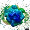
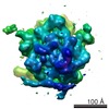
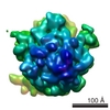
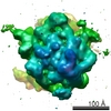
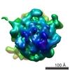
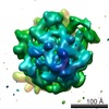
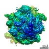
Cryo EM structure of E. coli 70S ribosome in complex with enzyme peptide deformylase and chaperone trigger factor
ボクセルのサイズ
X=Y=Z: 1.89 Å
密度
表面レベル
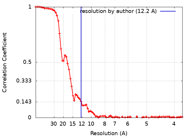
登録者による: 1.25 / ムービー #1: 1.25
最小 - 最大
-10.265516 - 13.369857
平均 (標準偏差)
0.06532132 (±1.1418496)
対称性
空間群: 1
詳細
EMDB XML:
マップ形状
Axis order
X
Y
Z
Origin
0
0
0
サイズ
220
220
220
Spacing
220
220
220
セル
A=B=C: 415.8 Å α=β=γ: 90.0 °
CCP4マップ ヘッダ情報:
mode
Image stored as Reals
Å/pix. X/Y/Z
1.89
1.89
1.89
M x/y/z
220
220
220
origin x/y/z
0.000
0.000
0.000
length x/y/z
415.800
415.800
415.800
α/β/γ
90.000
90.000
90.000
MAP C/R/S
1
2
3
start NC/NR/NS
0
0
0
NC/NR/NS
220
220
220
D min/max/mean
-10.266
13.370
0.065
-
添付データ
-
試料の構成要素
-
全体 : E. coli 70S ribosome in complex with enzyme peptide deformylase a...
全体
名称: E. coli 70S ribosome in complex with enzyme peptide deformylase and chaperone trigger factor
要素
複合体: E. coli 70S ribosome in complex with enzyme peptide deformylase and chaperone trigger factor
タンパク質・ペプチド: Peptide deformylase
タンパク質・ペプチド: Trigger factor
-
超分子 #1: E. coli 70S ribosome in complex with enzyme peptide deformylase a...
超分子
名称: E. coli 70S ribosome in complex with enzyme peptide deformylase and chaperone trigger factor タイプ: complex / ID: 1 / 親要素: 0 / 含まれる分子: all 詳細: The complex was prepared by incubating E. coli 70S ribosome with peptide deformylase, methionine aminopeptidase and trigger factor in that sequence. However, cryo EM reconstruction of the ...詳細: The complex was prepared by incubating E. coli 70S ribosome with peptide deformylase, methionine aminopeptidase and trigger factor in that sequence. However, cryo EM reconstruction of the complex showed no density corresponding to methionine aminopeptidase near the ribosomal tunnel exit.
 ムービー
ムービー コントローラー


 データを開く
 基本情報
基本情報 マップデータ
 試料
試料 キーワード
キーワード 機能・相同性情報
 データ登録者
 インド, 2件
  引用
引用 構造の表示
構造の表示
 ダウンロードとリンク
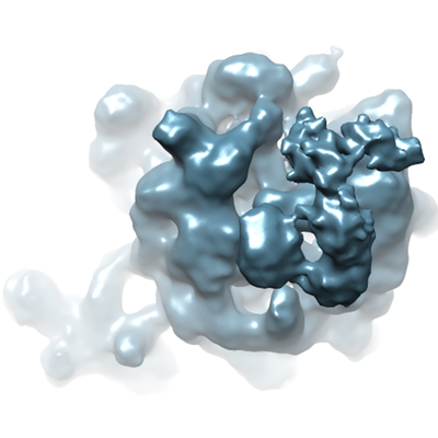 emd_9778.png
 http://ftp.pdbj.org/pub/emdb/structures/EMD-9778


 試料の構成要素
 解析
解析 電子顕微鏡法
 FIELD EMISSION GUN