Movie
Movie Controller

[English] 日本語
 Yorodumi
- PDB-6j45: Crystal structure of E. coli peptide deformylase enzyme and chape... -
+ Open data

- Basic information
| Entry | Database: PDB / ID: 6j45 | |||||||||
|---|---|---|---|---|---|---|---|---|---|---|
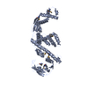
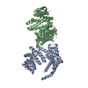
| Title | Crystal structure of E. coli peptide deformylase enzyme and chaperone trigger factor fitted into the cryo-EM density map of the complex | |||||||||
 Components Components |
| |||||||||
 Keywords Keywords | RIBOSOME / E. coli 70S ribosome / Protein biogenesis / Chaperone / Peptide deformylase / Trigger factor / Polypeptide exit tunnel / PPIase | |||||||||
| Function / homology |  Function and homology information Function and homology informationcell cycle / peptide deformylase / peptide deformylase activity / peptidylprolyl isomerase / peptidyl-prolyl cis-trans isomerase activity / protein transport / protein folding / translation / cell division / metal ion binding / cytoplasm Similarity search - Function | |||||||||
| Biological species |  | |||||||||
| Method | ELECTRON MICROSCOPY / single particle reconstruction / cryo EM / Resolution: 12.2 Å | |||||||||
 Authors Authors | Sengupta, J. / Bhakta, S. / Akbar, S. | |||||||||
| Funding support |  India, 2items India, 2items
| |||||||||
 Citation Citation | Journal: J Mol Biol / Year: 2019 Title: Cryo-EM Structures Reveal Relocalization of MetAP in the Presence of Other Protein Biogenesis Factors at the Ribosomal Tunnel Exit. Authors: Sayan Bhakta / Shirin Akbar / Jayati Sengupta / Abstract: During protein biosynthesis in bacteria, one of the earliest events that a nascent polypeptide chain goes through is the co-translational enzymatic processing. The event includes two enzymatic ...During protein biosynthesis in bacteria, one of the earliest events that a nascent polypeptide chain goes through is the co-translational enzymatic processing. The event includes two enzymatic pathways: deformylation of the N-terminal methionine by the enzyme peptide deformylase (PDF), followed by methionine excision catalyzed by methionine aminopeptidase (MetAP). During the enzymatic processing, the emerging nascent protein likely remains shielded by the ribosome-associated chaperone trigger factor. The ribosome tunnel exit serves as a stage for recruiting proteins involved in maturation processes of the nascent chain. Co-translational processing of nascent chains is a critical step for subsequent folding and functioning of mature proteins. Here, we present cryo-electron microscopy structures of Escherichia coli (E. coli) ribosome in complex with the nascent chain processing proteins. The structures reveal overlapping binding sites for PDF and MetAP when they bind individually at the tunnel exit site, where L22-L32 protein region provides primary anchoring sites for both proteins. In the absence of PDF, trigger factor can access ribosomal tunnel exit when MetAP occupies its primary binding site. Interestingly, however, in the presence of PDF, when MetAP's primary binding site is already engaged, MetAP has a remarkable ability to occupy an alternative binding site adjacent to PDF. Our study, thus, discloses an unexpected mechanism that MetAP adopts for context-specific ribosome association. | |||||||||
| History |
|
- Structure visualization
| Movie |
Movie viewer |
|---|---|
| Structure viewer | Molecule: MolmilJmol/JSmol |
- Downloads & links
-Download
| PDBx/mmCIF format | 6j45.cif.gz | 31.6 KB | Display | PDBx/mmCIF format |
|---|---|---|---|---|
| PDB format | pdb6j45.ent.gz | 14.5 KB | Display | PDB format |
| PDBx/mmJSON format | 6j45.json.gz | Tree view | PDBx/mmJSON format | |
| Others |  Other downloads Other downloads |
-Validation report
| Arichive directory | https://data.pdbj.org/pub/pdb/validation_reports/j4/6j45ftp://data.pdbj.org/pub/pdb/validation_reports/j4/6j45 | HTTPS FTP |
|---|
-Related structure data
| Related structure data | 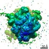 9778MC 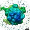 9750C 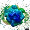 9752C  9753C  9759C  6iy7C 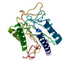 6iz7C 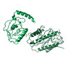 6iziC  6j0aC C: citing same article ( M: map data used to model this data |
|---|---|
| Similar structure data |


-Links
 PDBj
PDBj


- Assembly
| Deposited unit | 
|
|---|---|
| 1 |
|
-Components
| #1: Protein | Mass: 19357.447 Da / Num. of mol.: 1 Source method: isolated from a genetically manipulated source Source: (gene. exp.) |
|---|---|
| #2: Protein | Mass: 48255.570 Da / Num. of mol.: 1 Source method: isolated from a genetically manipulated source Source: (gene. exp.) |
-Experimental details
-Experiment
| Experiment | Method: ELECTRON MICROSCOPY |
|---|---|
| EM experiment | Aggregation state: PARTICLE / 3D reconstruction method: single particle reconstruction |
- Sample preparation
| Component | Name: E. coli 70S ribosome in complex with enzyme peptide deformylase and chaperone trigger factor Type: RIBOSOME Details: The complex was prepared by incubating E. coli 70S ribosome with peptide deformylase, methionine aminopeptidase and trigger factor in that sequence. However, cryo EM reconstruction of the ...Details: The complex was prepared by incubating E. coli 70S ribosome with peptide deformylase, methionine aminopeptidase and trigger factor in that sequence. However, cryo EM reconstruction of the complex showed no density corresponding to methionine aminopeptidase near the ribosomal tunnel exit. Entity ID: all / Source: MULTIPLE SOURCES | ||||||||||||
|---|---|---|---|---|---|---|---|---|---|---|---|---|---|
| Source (natural) |
| ||||||||||||
| Source (recombinant) |
| ||||||||||||
| Buffer solution | pH: 7.4 | ||||||||||||
| Specimen | Embedding applied: NO / Shadowing applied: NO / Staining applied: NO / Vitrification applied: YES | ||||||||||||
| Specimen support | Grid material: COPPER / Grid mesh size: 300 divisions/in. / Grid type: Quantifoil R2/2 | ||||||||||||
| Vitrification | Instrument: FEI VITROBOT MARK IV / Cryogen name: ETHANE |
- Electron microscopy imaging
| Experimental equipment |  Model: Tecnai Polara / Image courtesy: FEI Company |
|---|---|
| Microscopy | Model: FEI POLARA 300 |
| Electron gun | Electron source:  FIELD EMISSION GUN / Accelerating voltage: 300 kV / Illumination mode: FLOOD BEAM FIELD EMISSION GUN / Accelerating voltage: 300 kV / Illumination mode: FLOOD BEAM |
| Electron lens | Mode: BRIGHT FIELD |
| Specimen holder | Cryogen: NITROGEN |
| Image recording | Electron dose: 10 e/Å2 / Film or detector model: FEI EAGLE (4k x 4k) |
| Image scans | Width: 4096 / Height: 4096 |
- Processing
| EM software | Name: SPIDER / Category: 3D reconstruction | ||||||||||||
|---|---|---|---|---|---|---|---|---|---|---|---|---|---|
| CTF correction | Type: PHASE FLIPPING ONLY | ||||||||||||
| 3D reconstruction | Resolution: 12.2 Å / Resolution method: FSC 0.143 CUT-OFF / Num. of particles: 20900 / Symmetry type: POINT | ||||||||||||
| Atomic model building | 3D fitting-ID: 1 / Pdb chain-ID: A / Source name: PDB / Type: experimental model
|