Movie
Movie Controller

[English] 日本語
 Yorodumi
- EMDB-9318: Tetrahedral oligomeric complex of GyrA N-terminal fragment, solve... -
+ Open data

- Basic information
| Entry | Database: EMDB / ID: EMD-9318 | ||||||||||||||||||
|---|---|---|---|---|---|---|---|---|---|---|---|---|---|---|---|---|---|---|---|
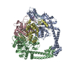
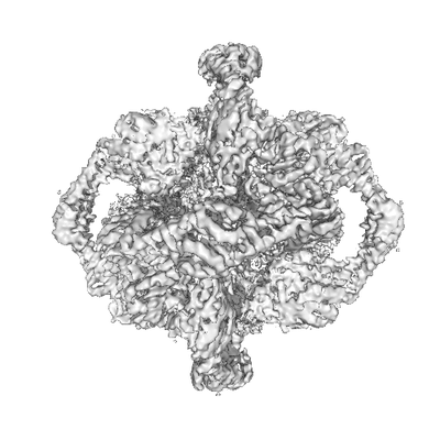
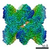
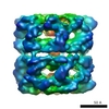
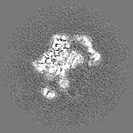
| Title | Tetrahedral oligomeric complex of GyrA N-terminal fragment, solved by cryoEM in tetrahedral symmetry | ||||||||||||||||||
 Map data Map data | Tetrahedral oligomeric complex of GyrA N-terminal fragment, solved by cryoEM in tetrahedral symmetry | ||||||||||||||||||
 Sample Sample |
| ||||||||||||||||||
 Keywords Keywords | topoisomerase / oligomeric complex / gyrase / ISOMERASE | ||||||||||||||||||
| Function / homology |  Function and homology information Function and homology informationDNA topoisomerase type II (double strand cut, ATP-hydrolyzing) complex / DNA negative supercoiling activity / DNA topoisomerase type II (double strand cut, ATP-hydrolyzing) activity / DNA topoisomerase (ATP-hydrolysing) / DNA topological change / DNA-templated DNA replication / chromosome / DNA binding / ATP binding / cytoplasm Similarity search - Function | ||||||||||||||||||
| Biological species |  Streptococcus pneumoniae G54 (bacteria) Streptococcus pneumoniae G54 (bacteria) | ||||||||||||||||||
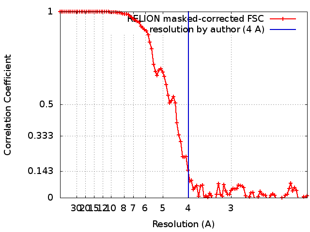
| Method | single particle reconstruction / cryo EM / Resolution: 4.0 Å | ||||||||||||||||||
 Authors Authors | Soczek KM / Grant T | ||||||||||||||||||
| Funding support |  United States, United States,  United Kingdom, 5 items United Kingdom, 5 items
| ||||||||||||||||||
 Citation Citation | Journal: Elife / Year: 2018 Title: CryoEM structures of open dimers of gyrase A in complex with DNA illuminate mechanism of strand passage. Authors: Katarzyna M Soczek / Tim Grant / Peter B Rosenthal / Alfonso Mondragón / Abstract: Gyrase is a unique type IIA topoisomerase that uses ATP hydrolysis to maintain the negatively supercoiled state of bacterial DNA. In order to perform its function, gyrase undergoes a sequence of ...Gyrase is a unique type IIA topoisomerase that uses ATP hydrolysis to maintain the negatively supercoiled state of bacterial DNA. In order to perform its function, gyrase undergoes a sequence of conformational changes that consist of concerted gate openings, DNA cleavage, and DNA strand passage events. Structures where the transported DNA molecule (T-segment) is trapped by the A subunit have not been observed. Here we present the cryoEM structures of two oligomeric complexes of open gyrase A dimers and DNA. The protein subunits in these complexes were solved to 4 Å and 5.2 Å resolution. One of the complexes traps a linear DNA molecule, a putative T-segment, which interacts with the open gyrase A dimers in two states, representing steps either prior to or after passage through the DNA-gate. The structures locate the T-segment in important intermediate conformations of the catalytic cycle and provide insights into gyrase-DNA interactions and mechanism. | ||||||||||||||||||
| History |
|
- Structure visualization
| Movie |
Movie viewer |
|---|---|
| Structure viewer | EM map: SurfViewMolmilJmol/JSmol |
| Supplemental images |
- Downloads & links
-EMDB archive
| Map data | emd_9318.map.gz | 73.3 MB | EMDB map data format | |
|---|---|---|---|---|
| Header (meta data) | emd-9318-v30.xmlemd-9318.xml | 16.5 KB 16.5 KB | Display Display | EMDB header |
| FSC (resolution estimation) | emd_9318_fsc.xml | 9.8 KB | Display | FSC data file |
| Images |  emd_9318.png emd_9318.png | 52.1 KB | ||
| Filedesc metadata | emd-9318.cif.gz | 6.4 KB | ||
| Archive directory |  http://ftp.pdbj.org/pub/emdb/structures/EMD-9318ftp://ftp.pdbj.org/pub/emdb/structures/EMD-9318 http://ftp.pdbj.org/pub/emdb/structures/EMD-9318ftp://ftp.pdbj.org/pub/emdb/structures/EMD-9318 | HTTPS FTP |
-Validation report
| Summary document | emd_9318_validation.pdf.gz | 605.6 KB | Display | EMDB validaton report |
|---|---|---|---|---|
| Full document | emd_9318_full_validation.pdf.gz | 605.2 KB | Display | |
| Data in XML | emd_9318_validation.xml.gz | 11.3 KB | Display | |
| Data in CIF | emd_9318_validation.cif.gz | 14.9 KB | Display | |
| Arichive directory | https://ftp.pdbj.org/pub/emdb/validation_reports/EMD-9318ftp://ftp.pdbj.org/pub/emdb/validation_reports/EMD-9318 | HTTPS FTP |
-Related structure data
| Related structure data |  6n1rMC  9316C 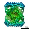 9317C 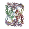 6n1pC 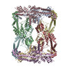 6n1qC C: citing same article ( M: atomic model generated by this map |
|---|---|
| Similar structure data |


-Links
| EMDB pages | EMDB (EBI/PDBe) / EMDataResource |
|---|---|
| Related items in Molecule of the Month |


-Map
| File | Download / File: emd_9318.map.gz / Format: CCP4 / Size: 78.5 MB / Type: IMAGE STORED AS FLOATING POINT NUMBER (4 BYTES) | ||||||||||||||||||||||||||||||||||||||||||||||||||||||||||||||||||||
|---|---|---|---|---|---|---|---|---|---|---|---|---|---|---|---|---|---|---|---|---|---|---|---|---|---|---|---|---|---|---|---|---|---|---|---|---|---|---|---|---|---|---|---|---|---|---|---|---|---|---|---|---|---|---|---|---|---|---|---|---|---|---|---|---|---|---|---|---|---|
| Annotation | Tetrahedral oligomeric complex of GyrA N-terminal fragment, solved by cryoEM in tetrahedral symmetry | ||||||||||||||||||||||||||||||||||||||||||||||||||||||||||||||||||||
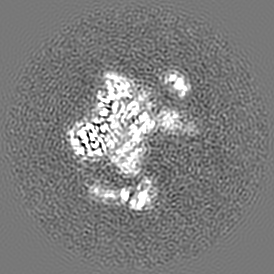
| Projections & slices | Image control
Images are generated by Spider. | ||||||||||||||||||||||||||||||||||||||||||||||||||||||||||||||||||||
| Voxel size | X=Y=Z: 1.04 Å | ||||||||||||||||||||||||||||||||||||||||||||||||||||||||||||||||||||
| Density |
| ||||||||||||||||||||||||||||||||||||||||||||||||||||||||||||||||||||
| Symmetry | Space group: 1 | ||||||||||||||||||||||||||||||||||||||||||||||||||||||||||||||||||||
| Details | EMDB XML:
CCP4 map header:
| ||||||||||||||||||||||||||||||||||||||||||||||||||||||||||||||||||||
 Z (Sec.)
 Y (Row.)
 X (Col.)


-Supplemental data
- Sample components
-Entire : Tetrahedral complex of DNA Gyrase A subunit N-terminal fragment, ...
| Entire | Name: Tetrahedral complex of DNA Gyrase A subunit N-terminal fragment, solved by cryoEM in T symmetry |
|---|---|
| Components |
|
-Supramolecule #1: Tetrahedral complex of DNA Gyrase A subunit N-terminal fragment, ...
| Supramolecule | Name: Tetrahedral complex of DNA Gyrase A subunit N-terminal fragment, solved by cryoEM in T symmetry type: complex / ID: 1 / Parent: 0 / Macromolecule list: all |
|---|---|
| Source (natural) | Organism: Streptococcus pneumoniae G54 (bacteria) |
| Molecular weight | Theoretical: 695 KDa |
-Macromolecule #1: DNA gyrase subunit A
| Macromolecule | Name: DNA gyrase subunit A / type: protein_or_peptide / ID: 1 / Number of copies: 12 / Enantiomer: LEVO / EC number: ec: 5.99.1.3 |
|---|---|
| Source (natural) | Organism: Streptococcus pneumoniae G54 (bacteria) |
| Molecular weight | Theoretical: 57.977578 KDa |
| Recombinant expression | Organism: |
| Sequence | String: MHHHHHHSSG VDLGTENLYF QSIAMQDKNL VNVNLTKEMK ASFIDYAMSV IVARALPDVR DGLKPVHRRI LYGMNELGVT PDKPHKKSA RITGDVMGKY HPHGDSSIYE AMVRMAQWWS YRYMLVDGHG NFGSMDGDSA AAQRYTEARM SKIALEMLRD I NKNTVDFV ...String: MHHHHHHSSG VDLGTENLYF QSIAMQDKNL VNVNLTKEMK ASFIDYAMSV IVARALPDVR DGLKPVHRRI LYGMNELGVT PDKPHKKSA RITGDVMGKY HPHGDSSIYE AMVRMAQWWS YRYMLVDGHG NFGSMDGDSA AAQRYTEARM SKIALEMLRD I NKNTVDFV DNYDANEREP LVLPARFPNL LVNGATGIAV GMATNIPPHN LGETIDAVKL VMDNPEVTTK DLMEVLPGPD FP TGALVMG KSGIHKAYET GKGSIVLRSR TEIETTKTGR ERIVVTEFPY MVNKTKVHEH IVRLVQEKRI EGITAVRDES NRE GVRFVI EVKRDASANV ILNNLFKMTQ MQTNFGFNML AIQNGIPKIL SLRQILDAYI EHQKEVVVRR TRFDKEKAEA RAHI LEGLL IALDHIDEVI RIIRASETDA EAQAELMSKF KLSERQSQAI LDMRLRRLTG LERDKIQSEY DDLLALIADL ADILA KPER VSQIIKDELD EVKRKFSDKR RTELMVG UniProtKB: DNA gyrase subunit A |
-Experimental details
-Structure determination
| Method | cryo EM |
|---|---|
 Processing Processing | single particle reconstruction |
| Aggregation state | particle |
-Sample preparation
| Buffer | pH: 7.5 Component:
| ||||||||||||
|---|---|---|---|---|---|---|---|---|---|---|---|---|---|
| Grid | Model: C-flat-1.2/1.3 4C / Material: COPPER / Mesh: 400 / Support film - Material: CARBON / Pretreatment - Type: GLOW DISCHARGE | ||||||||||||
| Vitrification | Cryogen name: ETHANE / Chamber humidity: 100 % / Chamber temperature: 277 K / Instrument: FEI VITROBOT MARK IV |
- Electron microscopy
| Microscope | FEI TITAN KRIOS |
|---|---|
| Temperature | Min: 100.0 K / Max: 100.0 K |
| Specialist optics | Energy filter - Name: GIF Quantum LS / Energy filter - Slit width: 20 eV |
| Image recording | Film or detector model: GATAN K2 SUMMIT (4k x 4k) / Detector mode: COUNTING / Digitization - Dimensions - Width: 3710 pixel / Digitization - Dimensions - Height: 3838 pixel / Digitization - Frames/image: 1-40 / Number real images: 4265 / Average exposure time: 14.0 sec. / Average electron dose: 74.0 e/Å2 |
| Electron beam | Acceleration voltage: 300 kV / Electron source:  FIELD EMISSION GUN FIELD EMISSION GUN |
| Electron optics | C2 aperture diameter: 50.0 µm / Calibrated magnification: 46430 / Illumination mode: OTHER / Imaging mode: BRIGHT FIELD / Cs: 2.7 mm / Nominal defocus max: 3.5 µm / Nominal defocus min: 1.0 µm / Nominal magnification: 130000 |
| Sample stage | Specimen holder model: FEI TITAN KRIOS AUTOGRID HOLDER / Cooling holder cryogen: NITROGEN |
| Experimental equipment |  Model: Titan Krios / Image courtesy: FEI Company |
+Image processing
-Atomic model buiding 1
| Initial model | PDB ID: Chain - Chain ID: A / Chain - Residue range: 2-486 / Chain - Source name: PDB / Chain - Initial model type: experimental model |
|---|---|
| Refinement | Space: REAL / Protocol: FLEXIBLE FIT / Target criteria: correlation coefficient |
| Output model | PDB-6n1r: |