large ribosomal subunit / ribosomal small subunit biogenesis / ribosomal small subunit assembly / small ribosomal subunit / small ribosomal subunit rRNA binding / transferase activity / 5S rRNA binding / large ribosomal subunit rRNA binding / cytosolic small ribosomal subunit / cytosolic large ribosomal subunit ...large ribosomal subunit / ribosomal small subunit biogenesis / ribosomal small subunit assembly / small ribosomal subunit / small ribosomal subunit rRNA binding / transferase activity / 5S rRNA binding / large ribosomal subunit rRNA binding / cytosolic small ribosomal subunit / cytosolic large ribosomal subunit / tRNA binding / cytoplasmic translation / rRNA binding / negative regulation of translation / ribosome / structural constituent of ribosome / ribonucleoprotein complex / translation / mRNA binding / RNA binding / zinc ion binding / cytoplasm / cytosol 類似検索 - 分子機能
Ribosomal protein L31 type B / Ribosomal protein L25, long-form / Ribosomal protein L25, beta domain / Ribosomal protein L25, C-terminal / Ribosomal protein TL5, C-terminal domain / Ribosomal protein S14, type Z / Ribosomal protein L31 signature. / Ribosomal protein L31 / Ribosomal protein L31 superfamily / Ribosomal protein L31 ...Ribosomal protein L31 type B / Ribosomal protein L25, long-form / Ribosomal protein L25, beta domain / Ribosomal protein L25, C-terminal / Ribosomal protein TL5, C-terminal domain / Ribosomal protein S14, type Z / Ribosomal protein L31 signature. / Ribosomal protein L31 / Ribosomal protein L31 superfamily / Ribosomal protein L31 / Ribosomal protein L16 signature 1. / : / Ribosomal protein L6, conserved site / Ribosomal protein L6 signature 1. / Ribosomal protein L16, conserved site / Ribosomal protein L16 signature 2. / Ribosomal protein L17 signature. / Ribosomal L25p family / Ribosomal protein L25 / Ribosomal protein L36 signature. / Ribosomal protein L28/L24 superfamily / Ribosomal protein L25/Gln-tRNA synthetase, N-terminal / Ribosomal protein L25/Gln-tRNA synthetase, anti-codon-binding domain superfamily / Ribosomal protein L28 / Ribosomal protein L35, conserved site / Ribosomal protein L35 signature. / Ribosomal protein L33, conserved site / Ribosomal protein L33 signature. / Ribosomal protein L35, non-mitochondrial / Ribosomal protein S14/S29 / Ribosomal protein L5, bacterial-type / Ribosomal protein L18, bacterial-type / Ribosomal protein L6, bacterial-type / Ribosomal protein L19, conserved site / Ribosomal protein L19 signature. / Ribosomal protein S3, bacterial-type / Ribosomal protein S6, conserved site / Ribosomal protein S6 signature. / Ribosomal protein L36 / Ribosomal protein L36 superfamily / Ribosomal protein L36 / Ribosomal protein S19, bacterial-type / Ribosomal protein L20 signature. / Ribosomal protein S7, bacterial/organellar-type / Ribosomal protein S11, bacterial-type / Ribosomal protein L27, conserved site / Ribosomal protein S13, bacterial-type / Ribosomal protein L27 signature. / Ribosomal protein S20 / Ribosomal protein S20 superfamily / Ribosomal protein S20 / Ribosomal protein S9, bacterial/plastid / Ribosomal protein S4, bacterial-type / 30S ribosomal protein S17 / Ribosomal protein S5, bacterial-type / Ribosomal protein L14P, bacterial-type / Ribosomal protein L34, conserved site / Ribosomal protein L34 signature. / Ribosomal protein S6, plastid/chloroplast / Ribosomal protein L22, bacterial/chloroplast-type / Ribosomal protein L2, bacterial/organellar-type / Ribosomal protein L35 / Ribosomal protein L35 superfamily / Ribosomal protein L35 / Ribosomal L28 family / Ribosomal protein L33 / Ribosomal protein L33 / Ribosomal protein L28/L24 / Ribosomal protein L18 / Ribosomal L18 of archaea, bacteria, mitoch. and chloroplast / Ribosomal protein L33 superfamily / Ribosomal protein L30, bacterial-type / : / Ribosomal protein L16 / Ribosomal protein S18, conserved site / Ribosomal protein S18 signature. / L28p-like / Ribosomal protein S16 / Ribosomal protein S16 / Ribosomal protein S16 domain superfamily / Ribosomal protein L20 / Ribosomal protein S15, bacterial-type / Ribosomal protein L20 / Ribosomal protein L20, C-terminal / Ribosomal protein L21 / Ribosomal protein L27 / Ribosomal L27 protein / Ribosomal protein L19 / Ribosomal protein L19 superfamily / Ribosomal protein L19 / Ribosomal proteins 50S L24/mitochondrial 39S L24 / Ribosomal protein L17 / Ribosomal protein L17 superfamily / Ribosomal protein L17 / Ribosomal protein S6 / Ribosomal protein S6 / Ribosomal protein S6 superfamily / Ribosomal protein L21-like / L21-like superfamily / Ribosomal prokaryotic L21 protein 類似検索 - ドメイン・相同性
50S ribosomal protein L32 / Large ribosomal subunit protein uL15 / Large ribosomal subunit protein uL30 / Small ribosomal subunit protein uS12 / Small ribosomal subunit protein uS7 / Large ribosomal subunit protein uL2 / Large ribosomal subunit protein bL34 / Large ribosomal subunit protein uL3 / Large ribosomal subunit protein uL4 / Large ribosomal subunit protein uL23 ...50S ribosomal protein L32 / Large ribosomal subunit protein uL15 / Large ribosomal subunit protein uL30 / Small ribosomal subunit protein uS12 / Small ribosomal subunit protein uS7 / Large ribosomal subunit protein uL2 / Large ribosomal subunit protein bL34 / Large ribosomal subunit protein uL3 / Large ribosomal subunit protein uL4 / Large ribosomal subunit protein uL23 / Small ribosomal subunit protein uS19 / Large ribosomal subunit protein uL22 / Small ribosomal subunit protein uS3 / Large ribosomal subunit protein uL16 / Large ribosomal subunit protein uL29 / Small ribosomal subunit protein uS17 / Large ribosomal subunit protein uL14 / Large ribosomal subunit protein uL24 / Large ribosomal subunit protein uL5 / Small ribosomal subunit protein uS14B / Small ribosomal subunit protein uS8 / Large ribosomal subunit protein uL6 / Large ribosomal subunit protein uL18 / Small ribosomal subunit protein uS5 / Large ribosomal subunit protein bL36 / Small ribosomal subunit protein uS13 / Small ribosomal subunit protein uS11 / Large ribosomal subunit protein bL17 / Large ribosomal subunit protein uL13 / Small ribosomal subunit protein uS9 / Large ribosomal subunit protein bL31B / Small ribosomal subunit protein uS4 / Large ribosomal subunit protein bL35 / Large ribosomal subunit protein bL20 / Large ribosomal subunit protein bL21 / Large ribosomal subunit protein bL27 / Small ribosomal subunit protein bS20 / Large ribosomal subunit protein bL33A / Large ribosomal subunit protein bL19 / Small ribosomal subunit protein bS16 / Large ribosomal subunit protein bL28 / Large ribosomal subunit protein bL25 / Small ribosomal subunit protein bS18 / Small ribosomal subunit protein bS6 / Small ribosomal subunit protein uS15 / 30S ribosomal protein S10 類似検索 - 構成要素
National Health and Medical Research Council (NHMRC, Australia)
1092262
オーストラリア
European Research Council (ERC)
322581 (NOVRIB)
European Union
引用
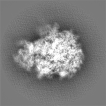
ジャーナル: mBio / 年: 2017 タイトル: Structural Basis for Linezolid Binding Site Rearrangement in the Ribosome. 著者: Matthew J Belousoff / Zohar Eyal / Mazdak Radjainia / Tofayel Ahmed / Rebecca S Bamert / Donna Matzov / Anat Bashan / Ella Zimmerman / Satabdi Mishra / David Cameron / Hans Elmlund / Anton Y ...著者: Matthew J Belousoff / Zohar Eyal / Mazdak Radjainia / Tofayel Ahmed / Rebecca S Bamert / Donna Matzov / Anat Bashan / Ella Zimmerman / Satabdi Mishra / David Cameron / Hans Elmlund / Anton Y Peleg / Shashi Bhushan / Trevor Lithgow / Ada Yonath / 要旨: An unorthodox, surprising mechanism of resistance to the antibiotic linezolid was revealed by cryo-electron microscopy (cryo-EM) in the 70S ribosomes from a clinical isolate of This high-resolution ...An unorthodox, surprising mechanism of resistance to the antibiotic linezolid was revealed by cryo-electron microscopy (cryo-EM) in the 70S ribosomes from a clinical isolate of This high-resolution structural information demonstrated that a single amino acid deletion in ribosomal protein uL3 confers linezolid resistance despite being located 24 Å away from the linezolid binding pocket in the peptidyl-transferase center. The mutation induces a cascade of allosteric structural rearrangements of the rRNA that ultimately results in the alteration of the antibiotic binding site. The growing burden on human health caused by various antibiotic resistance mutations now includes prevalent resistance to last-line antimicrobial drugs such as linezolid and daptomycin. Structure-informed drug modification represents a frontier with respect to designing advanced clinical therapies, but success in this strategy requires rapid, facile means to shed light on the structural basis for drug resistance (D. Brown, Nat Rev Drug Discov 14:821-832, 2015, https://doi.org/10.1038/nrd4675). Here, detailed structural information demonstrates that a common mechanism is at play in linezolid resistance and provides a step toward the redesign of oxazolidinone antibiotics, a strategy that could thwart known mechanisms of linezolid resistance.
 ムービー
ムービー コントローラー


 データを開く
 基本情報
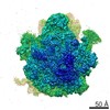
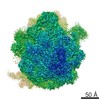
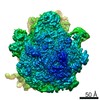
基本情報 マップデータ
 試料
試料 機能・相同性情報
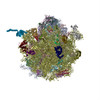
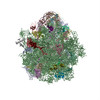
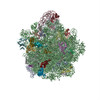
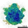
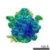
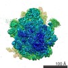
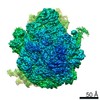
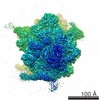
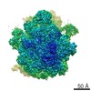
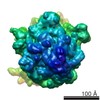
 Staphylococcus aureus (黄色ブドウ球菌) /
  データ登録者
 オーストラリア, European Union, 2件
  引用
引用

 構造の表示
構造の表示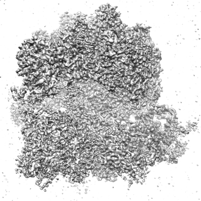
 ダウンロードとリンク
 emd_8402.png
 http://ftp.pdbj.org/pub/emdb/structures/EMD-8402
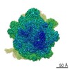


 Z (Sec.)
 Y (Row.)
 X (Col.)


 試料の構成要素
 解析
解析 電子顕微鏡法
 FIELD EMISSION GUN