 Movie
Movie Controller

[English] 日本語
 Yorodumi
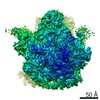
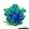
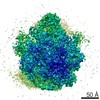
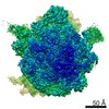
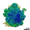
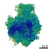
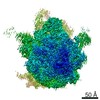
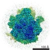
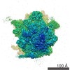
- PDB-5t7v: Methicillin Resistant, Linezolid resistant Staphylococcus aureus ... -
+ Open data

- Basic information
| Entry | Database: PDB / ID: 5t7v | ||||||||||||||||||||||||||||||||||||||||||||||||||||||||||||
|---|---|---|---|---|---|---|---|---|---|---|---|---|---|---|---|---|---|---|---|---|---|---|---|---|---|---|---|---|---|---|---|---|---|---|---|---|---|---|---|---|---|---|---|---|---|---|---|---|---|---|---|---|---|---|---|---|---|---|---|---|---|
| Title | Methicillin Resistant, Linezolid resistant Staphylococcus aureus 70S ribosome (delta S145 uL3) | ||||||||||||||||||||||||||||||||||||||||||||||||||||||||||||
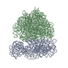
 Components Components |
| ||||||||||||||||||||||||||||||||||||||||||||||||||||||||||||
 Keywords Keywords | RIBOSOME / 70S ribosome / methicillin resistant / linezolid resistant / cryoEM | ||||||||||||||||||||||||||||||||||||||||||||||||||||||||||||
| Function / homology |  Function and homology information Function and homology informationlarge ribosomal subunit / transferase activity / ribosome biogenesis / ribosomal small subunit biogenesis / 5S rRNA binding / ribosomal large subunit assembly / small ribosomal subunit / small ribosomal subunit rRNA binding / large ribosomal subunit rRNA binding / cytosolic small ribosomal subunit ...large ribosomal subunit / transferase activity / ribosome biogenesis / ribosomal small subunit biogenesis / 5S rRNA binding / ribosomal large subunit assembly / small ribosomal subunit / small ribosomal subunit rRNA binding / large ribosomal subunit rRNA binding / cytosolic small ribosomal subunit / cytosolic large ribosomal subunit / cytoplasmic translation / tRNA binding / negative regulation of translation / rRNA binding / structural constituent of ribosome / ribosome / translation / ribonucleoprotein complex / mRNA binding / cytoplasm Similarity search - Function | ||||||||||||||||||||||||||||||||||||||||||||||||||||||||||||
| Biological species |   Staphylococcus aureus (bacteria) Staphylococcus aureus (bacteria) | ||||||||||||||||||||||||||||||||||||||||||||||||||||||||||||
| Method | ELECTRON MICROSCOPY / single particle reconstruction / cryo EM / Resolution: 3.6 Å | ||||||||||||||||||||||||||||||||||||||||||||||||||||||||||||
 Authors Authors | Belousoff, M.J. / Lithgow, T. / Eyal, Z. / Yonath, A. / Radjainia, M. | ||||||||||||||||||||||||||||||||||||||||||||||||||||||||||||
| Funding support |  Australia, 1items Australia, 1items
| ||||||||||||||||||||||||||||||||||||||||||||||||||||||||||||
 Citation Citation | Journal: mBio / Year: 2017 Title: Structural Basis for Linezolid Binding Site Rearrangement in the Ribosome. Authors: Matthew J Belousoff / Zohar Eyal / Mazdak Radjainia / Tofayel Ahmed / Rebecca S Bamert / Donna Matzov / Anat Bashan / Ella Zimmerman / Satabdi Mishra / David Cameron / Hans Elmlund / Anton Y ...Authors: Matthew J Belousoff / Zohar Eyal / Mazdak Radjainia / Tofayel Ahmed / Rebecca S Bamert / Donna Matzov / Anat Bashan / Ella Zimmerman / Satabdi Mishra / David Cameron / Hans Elmlund / Anton Y Peleg / Shashi Bhushan / Trevor Lithgow / Ada Yonath /   Abstract: An unorthodox, surprising mechanism of resistance to the antibiotic linezolid was revealed by cryo-electron microscopy (cryo-EM) in the 70S ribosomes from a clinical isolate of This high-resolution ...An unorthodox, surprising mechanism of resistance to the antibiotic linezolid was revealed by cryo-electron microscopy (cryo-EM) in the 70S ribosomes from a clinical isolate of This high-resolution structural information demonstrated that a single amino acid deletion in ribosomal protein uL3 confers linezolid resistance despite being located 24 Å away from the linezolid binding pocket in the peptidyl-transferase center. The mutation induces a cascade of allosteric structural rearrangements of the rRNA that ultimately results in the alteration of the antibiotic binding site. The growing burden on human health caused by various antibiotic resistance mutations now includes prevalent resistance to last-line antimicrobial drugs such as linezolid and daptomycin. Structure-informed drug modification represents a frontier with respect to designing advanced clinical therapies, but success in this strategy requires rapid, facile means to shed light on the structural basis for drug resistance (D. Brown, Nat Rev Drug Discov 14:821-832, 2015, https://doi.org/10.1038/nrd4675). Here, detailed structural information demonstrates that a common mechanism is at play in linezolid resistance and provides a step toward the redesign of oxazolidinone antibiotics, a strategy that could thwart known mechanisms of linezolid resistance. | ||||||||||||||||||||||||||||||||||||||||||||||||||||||||||||
| History |
|
- Structure visualization
| Movie |
Movie viewer |
|---|---|
| Structure viewer | Molecule: MolmilJmol/JSmol |
- Downloads & links
-Download
| PDBx/mmCIF format | 5t7v.cif.gz | 2.7 MB | Display | PDBx/mmCIF format |
|---|---|---|---|---|
| PDB format | pdb5t7v.ent.gz | Display | PDB format | |
| PDBx/mmJSON format | 5t7v.json.gz | Tree view | PDBx/mmJSON format | |
| Others |  Other downloads Other downloads |
-Validation report
| Arichive directory | https://data.pdbj.org/pub/pdb/validation_reports/t7/5t7vftp://data.pdbj.org/pub/pdb/validation_reports/t7/5t7v | HTTPS FTP |
|---|
-Related structure data
| Related structure data | 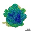 8369MC 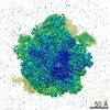 8402C 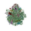 5tcuC M: map data used to model this data C: citing same article ( |
|---|---|
| Similar structure data |
-Links
 PDBj
PDBj


- Assembly
| Deposited unit | 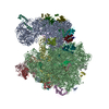
|
|---|---|
| 1 |
|
-Components
-RNA chain , 4 types, 4 molecules ABCD
| #1: RNA chain | Mass: 500520.406 Da / Num. of mol.: 1 / Source method: isolated from a natural source / Source: (natural) Staphylococcus aureus (bacteria) / References: GenBank: 1102759359 |
|---|---|
| #14: RNA chain | Mass: 945427.812 Da / Num. of mol.: 1 / Source method: isolated from a natural source / Source: (natural) Staphylococcus aureus (bacteria) / References: GenBank: 1015534143 |
| #15: RNA chain | Mass: 36652.777 Da / Num. of mol.: 1 / Source method: isolated from a natural source / Source: (natural) Staphylococcus aureus (bacteria) / References: GenBank: 1043615627 |
| #42: RNA chain | Mass: 23851.152 Da / Num. of mol.: 1 / Source method: isolated from a natural source / Source: (natural) Staphylococcus aureus (bacteria) |
-30S ribosomal protein ... , 12 types, 12 molecules S1S2S3S6S7S8S9SASCSDSESF
| #2: Protein | Mass: 9214.458 Da / Num. of mol.: 1 / Source method: isolated from a natural source / Source: (natural) Staphylococcus aureus (bacteria) / References: UniProt: A0A0H2K0A0 |
|---|---|
| #3: Protein | Mass: 12146.741 Da / Num. of mol.: 1 / Source method: isolated from a natural source / Source: (natural) Staphylococcus aureus (bacteria) / References: UniProt: A0A077UKD6 |
| #4: Protein | Mass: 15131.569 Da / Num. of mol.: 1 / Source method: isolated from a natural source / Source: (natural) Staphylococcus aureus (bacteria) / References: UniProt: W8U1C6 |
| #5: Protein | Mass: 10345.940 Da / Num. of mol.: 1 / Source method: isolated from a natural source / Source: (natural) Staphylococcus aureus (bacteria) / References: UniProt: W8U6H8 |
| #6: Protein | Mass: 8455.689 Da / Num. of mol.: 1 / Source method: isolated from a natural source / Source: (natural) Staphylococcus aureus (bacteria) / References: UniProt: A0A1K8TAS5 |
| #7: Protein | Mass: 9623.185 Da / Num. of mol.: 1 / Source method: isolated from a natural source / Source: (natural) Staphylococcus aureus (bacteria) / References: UniProt: A0A077VLD5 |
| #8: Protein | Mass: 6533.737 Da / Num. of mol.: 1 / Source method: isolated from a natural source / Source: (natural) Staphylococcus aureus (bacteria) / References: UniProt: A0A1D4K9U8 |
| #9: Protein | Mass: 8295.577 Da / Num. of mol.: 1 / Source method: isolated from a natural source / Source: (natural) Staphylococcus aureus (bacteria) / References: UniProt: A0A1K8EL93 |
| #10: Protein | Mass: 22763.027 Da / Num. of mol.: 1 / Source method: isolated from a natural source / Source: (natural) Staphylococcus aureus (bacteria) / References: UniProt: W8TVK2 |
| #11: Protein | Mass: 16245.786 Da / Num. of mol.: 1 / Source method: isolated from a natural source / Source: (natural) Staphylococcus aureus (bacteria) / References: UniProt: W8TUC9 |
| #12: Protein | Mass: 10864.370 Da / Num. of mol.: 1 / Source method: isolated from a natural source / Source: (natural) Staphylococcus aureus (bacteria) / References: UniProt: W8TPC6 |
| #13: Protein | Mass: 14536.908 Da / Num. of mol.: 1 / Source method: isolated from a natural source / Source: (natural) Staphylococcus aureus (bacteria) / References: UniProt: W8U8T8 |
+50S ribosomal protein ... , 26 types, 26 molecules L1L2L3L4L5L6L7L8L9LALBLCLDLELFLGLHLILJLLLMLNLOLPLQLR
-Non-polymers , 1 types, 17 molecules 
| #43: Chemical | ChemComp-MG /  Mass: 24.305 Da / Num. of mol.: 17 / Source method: obtained synthetically / Formula: Mg Mass: 24.305 Da / Num. of mol.: 17 / Source method: obtained synthetically / Formula: Mg |
|---|
-Details
| Has protein modification | Y |
|---|
-Experimental details
-Experiment
| Experiment | Method: ELECTRON MICROSCOPY |
|---|---|
| EM experiment | Aggregation state: PARTICLE / 3D reconstruction method: single particle reconstruction |
- Sample preparation
| Component | Name: Clinical isolate Linezolid resistant MRSA 70S ribosome Type: RIBOSOME / Entity ID: #1-#42 / Source: NATURAL |
|---|---|
| Molecular weight | Experimental value: NO |
| Source (natural) | Organism: Staphylococcus aureus (bacteria) |
| Buffer solution | pH: 7.5 |
| Specimen | Conc.: 0.3 mg/ml / Embedding applied: NO / Shadowing applied: NO / Staining applied: NO / Vitrification applied: YES |
| Specimen support | Grid material: COPPER / Grid mesh size: 300 divisions/in. / Grid type: Quantifoil R1.2/1.3 |
| Vitrification | Instrument: FEI VITROBOT MARK IV / Cryogen name: ETHANE / Humidity: 100 % / Chamber temperature: 277 K |
- Electron microscopy imaging
| Experimental equipment |  Model: Titan Krios / Image courtesy: FEI Company |
|---|---|
| Microscopy | Model: FEI TITAN KRIOS |
| Electron gun | Electron source:  FIELD EMISSION GUN / Accelerating voltage: 300 kV / Illumination mode: SPOT SCAN FIELD EMISSION GUN / Accelerating voltage: 300 kV / Illumination mode: SPOT SCAN |
| Electron lens | Mode: BRIGHT FIELD |
| Image recording | Electron dose: 45 e/Å2 / Film or detector model: FEI FALCON II (4k x 4k) |
- Processing
| Software | Name: PHENIX / Version: (dev_2429: phenix.real_space_refine) / Classification: refinement |
|---|---|
| EM software | Name: PHENIX / Category: model refinement |
| CTF correction | Type: PHASE FLIPPING AND AMPLITUDE CORRECTION |
| 3D reconstruction | Resolution: 3.6 Å / Resolution method: FSC 0.143 CUT-OFF / Num. of particles: 80500 / Symmetry type: POINT |
| Atomic model building | Protocol: FLEXIBLE FIT / Space: REAL |
