[English] 日本語
 Yorodumi
- EMDB-7291: Drosophila Dicer-2 apo homology model (helicase, Platform-PAZ, RN... -
+ Open data
- Basic information
| Entry | Database: EMDB / ID: EMD-7291 | |||||||||
|---|---|---|---|---|---|---|---|---|---|---|
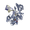
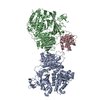
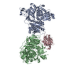
| Title | Drosophila Dicer-2 apo homology model (helicase, Platform-PAZ, RNaseIII domains) | |||||||||
 Map data Map data | Drosophila Dicer-2 (apo) | |||||||||
 Sample Sample |
| |||||||||
 Keywords Keywords | Dicer / dmDcr2 / Dicer-2 / helicase / platform / PAZ / RNAseIII / RNA BINDING PROTEIN | |||||||||
| Function / homology |  Function and homology information Function and homology informationpositive regulation of Toll signaling pathway / lncRNA catabolic process / RNAi-mediated antiviral immune response / dosage compensation by hyperactivation of X chromosome / global gene silencing by mRNA cleavage / apoptotic DNA fragmentation / ribonuclease III / deoxyribonuclease I activity / RISC-loading complex / detection of virus ...positive regulation of Toll signaling pathway / lncRNA catabolic process / RNAi-mediated antiviral immune response / dosage compensation by hyperactivation of X chromosome / global gene silencing by mRNA cleavage / apoptotic DNA fragmentation / ribonuclease III / deoxyribonuclease I activity / RISC-loading complex / detection of virus / RISC complex assembly / regulatory ncRNA-mediated post-transcriptional gene silencing / ribonuclease III activity / siRNA processing / siRNA binding / ATP-dependent activity, acting on RNA / RISC complex / positive regulation of innate immune response / positive regulation of defense response to virus by host / mRNA 3'-UTR binding / locomotory behavior / helicase activity / cellular response to virus / cytoplasmic ribonucleoprotein granule / heterochromatin formation / defense response to virus / perinuclear region of cytoplasm / ATP hydrolysis activity / RNA binding / ATP binding / nucleus / cytoplasm Similarity search - Function | |||||||||
| Biological species |  | |||||||||
| Method | single particle reconstruction / cryo EM / Resolution: 7.1 Å | |||||||||
 Authors Authors | Shen PS / Sinha NK | |||||||||
| Funding support |  United States, 1 items United States, 1 items
| |||||||||
 Citation Citation |  Journal: Science / Year: 2018 Journal: Science / Year: 2018Title: Dicer uses distinct modules for recognizing dsRNA termini. Authors: Niladri K Sinha / Janet Iwasa / Peter S Shen / Brenda L Bass /  Abstract: Invertebrates rely on Dicer to cleave viral double-stranded RNA (dsRNA), and Dicer-2 distinguishes dsRNA substrates by their termini. Blunt termini promote processive cleavage, while 3' overhanging ...Invertebrates rely on Dicer to cleave viral double-stranded RNA (dsRNA), and Dicer-2 distinguishes dsRNA substrates by their termini. Blunt termini promote processive cleavage, while 3' overhanging termini are cleaved distributively. To understand this discrimination, we used cryo-electron microscopy to solve structures of Dicer-2 alone and in complex with blunt dsRNA. Whereas the Platform-PAZ domains have been considered the only Dicer domains that bind dsRNA termini, unexpectedly, we found that the helicase domain is required for binding blunt, but not 3' overhanging, termini. We further showed that blunt dsRNA is locally unwound and threaded through the helicase domain in an adenosine triphosphate-dependent manner. Our studies reveal a previously unrecognized mechanism for optimizing antiviral defense and set the stage for the discovery of helicase-dependent functions in other Dicers. | |||||||||
| History |
|
- Structure visualization
| Movie |
 Movie viewer Movie viewer |
|---|---|
| Structure viewer | EM map:  SurfView SurfView Molmil Molmil Jmol/JSmol Jmol/JSmol |
| Supplemental images |
- Downloads & links
-EMDB archive
| Map data |  emd_7291.map.gz emd_7291.map.gz | 49.2 MB |  EMDB map data format EMDB map data format | |
|---|---|---|---|---|
| Header (meta data) |  emd-7291-v30.xml emd-7291-v30.xml emd-7291.xml emd-7291.xml | 11 KB 11 KB | Display Display |  EMDB header EMDB header |
| Images |  emd_7291.png emd_7291.png | 32.5 KB | ||
| Filedesc metadata |  emd-7291.cif.gz emd-7291.cif.gz | 6.1 KB | ||
| Archive directory |  http://ftp.pdbj.org/pub/emdb/structures/EMD-7291 http://ftp.pdbj.org/pub/emdb/structures/EMD-7291 ftp://ftp.pdbj.org/pub/emdb/structures/EMD-7291 ftp://ftp.pdbj.org/pub/emdb/structures/EMD-7291 | HTTPS FTP |
-Related structure data
| Related structure data |  6buaMC  7290C  7292C  6bu9C C: citing same article ( M: atomic model generated by this map |
|---|---|
| Similar structure data |
- Links
Links
| EMDB pages |  EMDB (EBI/PDBe) / EMDB (EBI/PDBe) /  EMDataResource EMDataResource |
|---|---|
| Related items in Molecule of the Month |
- Map
Map
| File |  Download / File: emd_7291.map.gz / Format: CCP4 / Size: 64 MB / Type: IMAGE STORED AS FLOATING POINT NUMBER (4 BYTES) Download / File: emd_7291.map.gz / Format: CCP4 / Size: 64 MB / Type: IMAGE STORED AS FLOATING POINT NUMBER (4 BYTES) | ||||||||||||||||||||||||||||||||||||||||||||||||||||||||||||
|---|---|---|---|---|---|---|---|---|---|---|---|---|---|---|---|---|---|---|---|---|---|---|---|---|---|---|---|---|---|---|---|---|---|---|---|---|---|---|---|---|---|---|---|---|---|---|---|---|---|---|---|---|---|---|---|---|---|---|---|---|---|
| Annotation | Drosophila Dicer-2 (apo) | ||||||||||||||||||||||||||||||||||||||||||||||||||||||||||||
| Projections & slices | Image control
Images are generated by Spider. | ||||||||||||||||||||||||||||||||||||||||||||||||||||||||||||
| Voxel size | X=Y=Z: 1.193 Å | ||||||||||||||||||||||||||||||||||||||||||||||||||||||||||||
| Density |
| ||||||||||||||||||||||||||||||||||||||||||||||||||||||||||||
| Symmetry | Space group: 1 | ||||||||||||||||||||||||||||||||||||||||||||||||||||||||||||
| Details | EMDB XML:
CCP4 map header:
| ||||||||||||||||||||||||||||||||||||||||||||||||||||||||||||
-Supplemental data
- Sample components
-Entire : Drosophila Dicer-2 (apo)
| Entire | Name: Drosophila Dicer-2 (apo) |
|---|---|
| Components |
|
-Supramolecule #1: Drosophila Dicer-2 (apo)
| Supramolecule | Name: Drosophila Dicer-2 (apo) / type: complex / ID: 1 / Parent: 0 / Macromolecule list: all |
|---|---|
| Source (natural) | Organism:  |
-Macromolecule #1: Dicer-2, isoform A
| Macromolecule | Name: Dicer-2, isoform A / type: protein_or_peptide / ID: 1 / Number of copies: 1 / Enantiomer: LEVO / EC number: ribonuclease III |
|---|---|
| Source (natural) | Organism:  |
| Molecular weight | Theoretical: 198.074797 KDa |
| Recombinant expression | Organism:  |
| Sequence | String: GPMEDVEIKP RGYQLRLVDH LTKSNGIVYL PTGSGKTFVA ILVLKRFSQD FDKPIESGGK RALFMCNTVE LARQQAMAVR RCTNFKVGF YVGEQGVDDW TRGMWSDEIK KNQVLVGTAQ VFLDMVTQTY VALSSLSVVI IDECHHGTGH HPFREFMRLF T IANQTKLP ...String: GPMEDVEIKP RGYQLRLVDH LTKSNGIVYL PTGSGKTFVA ILVLKRFSQD FDKPIESGGK RALFMCNTVE LARQQAMAVR RCTNFKVGF YVGEQGVDDW TRGMWSDEIK KNQVLVGTAQ VFLDMVTQTY VALSSLSVVI IDECHHGTGH HPFREFMRLF T IANQTKLP RVVGLTGVLI KGNEITNVAT KLKELEITYR GNIITVSDTK EMENVMLYAT KPTEVMVSFP HQEQVLTVTR LI SAEIEKF YVSLDLMNIG VQPIRRSKSL QCLRDPSKKS FVKQLFNDFL YQMKEYGIYA ASIAIISLIV EFDIKRRQAE TLS VKLMHR TALTLCEKIR HLLVQKLQDM TYDDDDDNVN TEEVIMNFST PKVQRFLMSL KVSFADKDPK DICCLVFVER RYTC KCIYG LLLNYIQSTP ELRNVLTPQF MVGRNNISPD FESVLERKWQ KSAIQQFRDG NANLMICSSV LEEGIDVQAC NHVFI LDPV KTFNMYVQSK GRARTTEAKF VLFTADKERE KTIQQIYQYR KAHNDIAEYL KDRVLEKTEP ELYEIKGHFQ DDIDPF TNE NGAVLLPNNA LAILHRYCQT IPTDAFGFVI PWFHVLQEDE RDRIFGVSAK GKHVISINMP VNCMLRDTIY SDPMDNV KT AKISAAFKAC KVLYSLGELN ERFVPKTLKE RVASIADVHF EHWNKYGDSV TATVNKADKS KDRTYKTECP LEFYDALP R VGEICYAYEI FLEPQFESCE YTEHMYLNLQ TPRNYAILLR NKLPRLAEMP LFSNQGKLHV RVANAPLEVI IQNSEQLEL LHQFHGMVFR DILKIWHPFF VLDRRSKENS YLVVPLILGA GEQKCFDWEL MTNFRRLPQS HGSNVQQREQ QPAPRPEDFE GKIVTQWYA NYDKPMLVTK VHRELTPLSY MEKNQQDKTY YEFTMSKYGN RIGDVVHKDK FMIEVRDLTE QLTFYVHNRG K FNAKSKAK MKVILIPELC FNFNFPGDLW LKLIFLPSIL NRMYFLLHAE ALRKRFNTYL NLHLLPFNGT DYMPRPLEID YS LKRNVDP LGNVIPTEDI EEPKSLLEPM PTKSIEASVA NLEITEFENP WQKYMEPVDL SRNLLSTYPV ELDYYYHFSV GNV CEMNEM DFEDKEYWAK NQFHMPTGNI YGNRTPAKTN ANVPALMPSK PTVRGKVKPL LILQKTVSKE HITPAEQGEF LAAI TASSA ADVFDMERLE ILGASFLKLS ATLYLASKYS DWNEGTLTEV KSKLVSNRNL LFCLIDADIP KTLNTIQFTP RYTWL PPGI SLPHNVLALW RENPEFAKII GPHNLRDLAL GDEESLVKGN CSDINYNRFV EGCRANGQSF YAGADFSSEV NFCVGL VTI PNKVIADTLE ALLGVIVKNY GLQHAFKMLE YFKICRADID KPLTQLLNLE LGGKKMRANV NTTEIDGFLI NHYYLEK NL GYTFKDRRYL LQALTHPSYP TNRITGSYQE LEFIGAAILD FLISAYIFEN NTKMNPGALT DLRSALVNNT TLACICVR H RLHFFILAEN AKLSEIISKF VNFQESQGHR VTNYVRILLE EADVQPTPLD LDDELDMTEL PHANKCISQE AEKGVPPKG EFNMSTNVDV PKALGDVLEA LIAAVYLDCR DLQRTWEVIF NLFEPELQEF TRKVPINHIR QLVEHKHAKP VFSSPIVEGE TVMVSCQFT CMEKTIKVYG FGSNKDQAKL SAAKHALQQL SKCDA UniProtKB: Endoribonuclease Dcr-2 |
-Experimental details
-Structure determination
| Method | cryo EM |
|---|---|
 Processing Processing | single particle reconstruction |
| Aggregation state | particle |
- Sample preparation
| Buffer | pH: 8 |
|---|---|
| Vitrification | Cryogen name: ETHANE |
- Electron microscopy
| Microscope | FEI TECNAI F20 |
|---|---|
| Image recording | Film or detector model: GATAN K2 SUMMIT (4k x 4k) / Average electron dose: 1.2 e/Å2 |
| Electron beam | Acceleration voltage: 200 kV / Electron source:  FIELD EMISSION GUN FIELD EMISSION GUN |
| Electron optics | Illumination mode: FLOOD BEAM / Imaging mode: BRIGHT FIELD |
| Experimental equipment |  Model: Tecnai F20 / Image courtesy: FEI Company |
- Image processing
| Startup model | Type of model: PDB ENTRY PDB model - PDB ID: |
|---|---|
| Final reconstruction | Resolution.type: BY AUTHOR / Resolution: 7.1 Å / Resolution method: FSC 0.143 CUT-OFF / Number images used: 85119 |
| Initial angle assignment | Type: RANDOM ASSIGNMENT |
| Final angle assignment | Type: ANGULAR RECONSTITUTION |
 Movie
Movie Controller




 Z (Sec.)
 Y (Row.)
 X (Col.)


