 Movie
Movie Controller


+ Open data

- Basic information
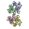
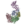
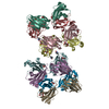
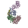
| Entry | Database: EMDB / ID: EMD-7292 | |||||||||
|---|---|---|---|---|---|---|---|---|---|---|
| Title | Drosophila Dicer-2 (apo, intact helicase density) | |||||||||
 Map data Map data | Drosophila Dicer-2 (intact helicase density) | |||||||||
 Sample Sample |
| |||||||||
| Biological species |  | |||||||||
| Method | single particle reconstruction / cryo EM / Resolution: 8.7 Å | |||||||||
 Authors Authors | Shen PS / Sinha NK / Bass BL | |||||||||
 Citation Citation | Journal: Science / Year: 2018 Title: Dicer uses distinct modules for recognizing dsRNA termini. Authors: Niladri K Sinha / Janet Iwasa / Peter S Shen / Brenda L Bass /  Abstract: Invertebrates rely on Dicer to cleave viral double-stranded RNA (dsRNA), and Dicer-2 distinguishes dsRNA substrates by their termini. Blunt termini promote processive cleavage, while 3' overhanging ...Invertebrates rely on Dicer to cleave viral double-stranded RNA (dsRNA), and Dicer-2 distinguishes dsRNA substrates by their termini. Blunt termini promote processive cleavage, while 3' overhanging termini are cleaved distributively. To understand this discrimination, we used cryo-electron microscopy to solve structures of Dicer-2 alone and in complex with blunt dsRNA. Whereas the Platform-PAZ domains have been considered the only Dicer domains that bind dsRNA termini, unexpectedly, we found that the helicase domain is required for binding blunt, but not 3' overhanging, termini. We further showed that blunt dsRNA is locally unwound and threaded through the helicase domain in an adenosine triphosphate-dependent manner. Our studies reveal a previously unrecognized mechanism for optimizing antiviral defense and set the stage for the discovery of helicase-dependent functions in other Dicers. | |||||||||
| History |
|
- Structure visualization
| Movie |
 Movie viewer Movie viewer |
|---|---|
| Structure viewer | EM map: SurfViewMolmilJmol/JSmol |
| Supplemental images |

- Downloads & links
-EMDB archive
| Map data | emd_7292.map.gz | 8.9 MB | EMDB map data format | |
|---|---|---|---|---|
| Header (meta data) | emd-7292-v30.xmlemd-7292.xml | 10.4 KB 10.4 KB | Display Display | EMDB header |
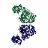
| Images | 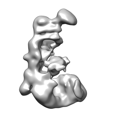 emd_7292.png emd_7292.png | 39.4 KB | ||
| Archive directory |  http://ftp.pdbj.org/pub/emdb/structures/EMD-7292ftp://ftp.pdbj.org/pub/emdb/structures/EMD-7292 http://ftp.pdbj.org/pub/emdb/structures/EMD-7292ftp://ftp.pdbj.org/pub/emdb/structures/EMD-7292 | HTTPS FTP |
-Related structure data


-Links
| EMDB pages | EMDB (EBI/PDBe) / EMDataResource |
|---|
-Map
| File | Download / File: emd_7292.map.gz / Format: CCP4 / Size: 64 MB / Type: IMAGE STORED AS FLOATING POINT NUMBER (4 BYTES) | ||||||||||||||||||||||||||||||||||||||||||||||||||||||||||||||||||||
|---|---|---|---|---|---|---|---|---|---|---|---|---|---|---|---|---|---|---|---|---|---|---|---|---|---|---|---|---|---|---|---|---|---|---|---|---|---|---|---|---|---|---|---|---|---|---|---|---|---|---|---|---|---|---|---|---|---|---|---|---|---|---|---|---|---|---|---|---|---|
| Annotation | Drosophila Dicer-2 (intact helicase density) | ||||||||||||||||||||||||||||||||||||||||||||||||||||||||||||||||||||
| Projections & slices | Image control
Images are generated by Spider. | ||||||||||||||||||||||||||||||||||||||||||||||||||||||||||||||||||||
| Voxel size | X=Y=Z: 1.193 Å | ||||||||||||||||||||||||||||||||||||||||||||||||||||||||||||||||||||
| Density |
| ||||||||||||||||||||||||||||||||||||||||||||||||||||||||||||||||||||
| Symmetry | Space group: 1 | ||||||||||||||||||||||||||||||||||||||||||||||||||||||||||||||||||||
| Details | EMDB XML:
CCP4 map header:
| ||||||||||||||||||||||||||||||||||||||||||||||||||||||||||||||||||||
 Z (Sec.)
 Y (Row.)
 X (Col.)


-Supplemental data
- Sample components
-Entire : Drosophila Dicer-2 (apo)
| Entire | Name: Drosophila Dicer-2 (apo) |
|---|---|
| Components |
|
-Supramolecule #1: Drosophila Dicer-2 (apo)
| Supramolecule | Name: Drosophila Dicer-2 (apo) / type: complex / ID: 1 / Parent: 0 / Macromolecule list: all |
|---|---|
| Source (natural) | Organism: |
| Recombinant expression | Organism:  Spodoptera frugiperda (fall armyworm) Spodoptera frugiperda (fall armyworm) |
-Macromolecule #1: Dicer-2
| Macromolecule | Name: Dicer-2 / type: protein_or_peptide / ID: 1 / Enantiomer: LEVO |
|---|---|
| Source (natural) | Organism: |
| Recombinant expression | Organism: Spodoptera frugiperda (fall armyworm) |
| Sequence | String: GPMEDVEIKP RGYQLRLVDH LTKSNGIVYL PTGSGKTFVA ILVLKRFSQD FDKPIESGGK RALFMCNTVE LARQQAMAVR RCTNFKVGFY VGEQGVDDWT RGMWSDEIKK NQVLVGTAQV FLDMVTQTYV ALSSLSVVII DECHHGTGHH PFREFMRLFT IANQTKLPRV ...String: GPMEDVEIKP RGYQLRLVDH LTKSNGIVYL PTGSGKTFVA ILVLKRFSQD FDKPIESGGK RALFMCNTVE LARQQAMAVR RCTNFKVGFY VGEQGVDDWT RGMWSDEIKK NQVLVGTAQV FLDMVTQTYV ALSSLSVVII DECHHGTGHH PFREFMRLFT IANQTKLPRV VGLTGVLIKG NEITNVATKL KELEITYRGN IITVSDTKEM ENVMLYATKP TEVMVSFPHQ EQVLTVTRLI SAEIEKFYVS LDLMNIGVQP IRRSKSLQCL RDPSKKSFVK QLFNDFLYQM KEYGIYAASI AIISLIVEFD IKRRQAETLS VKLMHRTALT LCEKIRHLLV QKLQDMTYDD DDDNVNTEEV IMNFSTPKVQ RFLMSLKVSF ADKDPKDICC LVFVERRYTC KCIYGLLLNY IQSTPELRNV LTPQFMVGRN NISPDFESVL ERKWQKSAIQ QFRDGNANLM ICSSVLEEGI DVQACNHVFI LDPVKTFNMY VQSKGRARTT EAKFVLFTAD KEREKTIQQI YQYRKAHNDI AEYLKDRVLE KTEPELYEIK GHFQDDIDPF TNENGAVLLP NNALAILHRY CQTIPTDAFG FVIPWFHVLQ EDERDRIFGV SAKGKHVISI NMPVNCMLRD TIYSDPMDNV KTAKISAAFK ACKVLYSLGE LNERFVPKTL KERVASIADV HFEHWNKYGD SVTATVNKAD KSKDRTYKTE CPLEFYDALP RVGEICYAYE IFLEPQFESC EYTEHMYLNL QTPRNYAILL RNKLPRLAEM PLFSNQGKLH VRVANAPLEV IIQNSEQLEL LHQFHGMVFR DILKIWHPFF VLDRRSKENS YLVVPLILGA GEQKCFDWEL MTNFRRLPQS HGSNVQQREQ QPAPRPEDFE GKIVTQWYAN YDKPMLVTKV HRELTPLSYM EKNQQDKTYY EFTMSKYGNR IGDVVHKDKF MIEVRDLTEQ LTFYVHNRGK FNAKSKAKMK VILIPELCFN FNFPGDLWLK LIFLPSILNR MYFLLHAEAL RKRFNTYLNL HLLPFNGTDY MPRPLEIDYS LKRNVDPLGN VIPTEDIEEP KSLLEPMPTK SIEASVANLE ITEFENPWQK YMEPVDLSRN LLSTYPVELD YYYHFSVGNV CEMNEMDFED KEYWAKNQFH MPTGNIYGNR TPAKTNANVP ALMPSKPTVR GKVKPLLILQ KTVSKEHITP AEQGEFLAAI TASSAADVFD MERLEILGAS FLKLSATLYL ASKYSDWNEG TLTEVKSKLV SNRNLLFCLI DADIPKTLNT IQFTPRYTWL PPGISLPHNV LALWRENPEF AKIIGPHNLR DLALGDEESL VKGNCSDINY NRFVEGCRAN GQSFYAGADF SSEVNFCVGL VTIPNKVIAD TLEALLGVIV KNYGLQHAFK MLEYFKICRA DIDKPLTQLL NLELGGKKMR ANVNTTEIDG FLINHYYLEK NLGYTFKDRR YLLQALTHPS YPTNRITGSY QELEFIGAAI LDFLISAYIF ENNTKMNPGA LTDLRSALVN NTTLACICVR HRLHFFILAE NAKLSEIISK FVNFQESQGH RVTNYVRILL EEADVQPTPL DLDDELDMTE LPHANKCISQ EAEKGVPPKG EFNMSTNVDV PKALGDVLEA LIAAVYLDCR DLQRTWEVIF NLFEPELQEF TRKVPINHIR QLVEHKHAKP VFSSPIVEGE TVMVSCQFTC MEKTIKVYGF GSNKDQAKLS AAKHALQQLS KCDA |
-Experimental details
-Structure determination
| Method | cryo EM |
|---|---|
 Processing Processing | single particle reconstruction |
| Aggregation state | particle |
-Sample preparation
| Buffer | pH: 8 |
|---|---|
| Vitrification | Cryogen name: ETHANE |
- Electron microscopy
| Microscope | FEI TECNAI F20 |
|---|---|
| Image recording | Film or detector model: GATAN K2 SUMMIT (4k x 4k) / Average electron dose: 1.2 e/Å2 |
| Electron beam | Acceleration voltage: 200 kV / Electron source:  FIELD EMISSION GUN FIELD EMISSION GUN |
| Electron optics | Illumination mode: FLOOD BEAM / Imaging mode: BRIGHT FIELD |
| Experimental equipment |  Model: Tecnai F20 / Image courtesy: FEI Company |
-Image processing
| Final reconstruction | Applied symmetry - Point group: C1 (asymmetric) / Resolution.type: BY AUTHOR / Resolution: 8.7 Å / Resolution method: FSC 0.143 CUT-OFF / Number images used: 9730 |
|---|---|
| Initial angle assignment | Type: RANDOM ASSIGNMENT |
| Final angle assignment | Type: ANGULAR RECONSTITUTION |
