National Institutes of Health/National Institute of General Medical Sciences (NIH/NIGMS)
R35 GM150960
United States
Citation
Journal: J Struct Biol / Year: 2025 Title: Nickel-NTA lipid-monolayer affinity grids allow for high-resolution structure determination by cryo-EM. Authors: Aleksandra Skrajna / Clara Lenger / Emily Robinson / Kevin Cannon / Reta Sarsam / Richard G Ouellette / Alberta M Abotsi / Patrick Brennwald / Robert K McGinty / Joshua D Strauss / Richard W Baker / Abstract: Grid preparation is a rate-limiting step in determining high-resolution structures by single particle cryo-EM. Particle interaction with the air-water interface often leads to denaturation, ...Grid preparation is a rate-limiting step in determining high-resolution structures by single particle cryo-EM. Particle interaction with the air-water interface often leads to denaturation, aggregation, or a preferred orientation within the ice. Some samples yield insufficient quantities of particles when using traditional grid making techniques and require the use of solid supports that concentrate samples onto the grid. Recent advances in grid-preparation show that affinity grids are promising tools to selectively concentrate proteins while simultaneously protecting samples from the air-water interface. One such technique utilizes lipid monolayers containing a lipid species with an affinity handle. Some of the first affinity grids used a holey carbon layer coated with nickel nitrilotriacetic acid (Ni-NTA) lipid, which allowed for the binding of proteins bearing the commonly used poly-histidine affinity tag. These studies however used complicated protocols and were conducted before the "resolution revolution" of cryo-EM. Here, we provide a straightforward preparation method and systematic analysis of Ni-NTA lipid monolayers as a tool for high-resolution single particle cryo-EM. We found the lipid affinity grids concentrate particles away from the AWI in thin ice (∼30 nm). We determined three structures ranging from 2.4 to 3.0 Å resolution, showing this method is amenable to high-resolution. Furthermore, we determined a 3.1 Å structure of a sub-100 kDa protein without symmetry, demonstrating the utility for a range of biological macromolecules. Lipid monolayers are therefore an easily extendable tool for most systems and help alleviate common problems such as low yield, disruption by the air-water interface, and thicker ice.
In the structure databanks used in Yorodumi, some data are registered as the other names, "COVID-19 virus" and "2019-nCoV". Here are the details of the virus and the list of structure data.
Jan 31, 2019. EMDB accession codes are about to change! (news from PDBe EMDB page)
EMDB accession codes are about to change! (news from PDBe EMDB page)
The allocation of 4 digits for EMDB accession codes will soon come to an end. Whilst these codes will remain in use, new EMDB accession codes will include an additional digit and will expand incrementally as the available range of codes is exhausted. The current 4-digit format prefixed with “EMD-” (i.e. EMD-XXXX) will advance to a 5-digit format (i.e. EMD-XXXXX), and so on. It is currently estimated that the 4-digit codes will be depleted around Spring 2019, at which point the 5-digit format will come into force.
The EM Navigator/Yorodumi systems omit the EMD- prefix.
Related info.:Q: What is EMD? / ID/Accession-code notation in Yorodumi/EM Navigator
Yorodumi is a browser for structure data from EMDB, PDB, SASBDB, etc.
This page is also the successor to EM Navigator detail page, and also detail information page/front-end page for Omokage search.
The word "yorodu" (or yorozu) is an old Japanese word meaning "ten thousand". "mi" (miru) is to see.
Related info.:EMDB / PDB / SASBDB / Comparison of 3 databanks / Yorodumi Search / Aug 31, 2016. New EM Navigator & Yorodumi / Yorodumi Papers / Jmol/JSmol / Function and homology information / Changes in new EM Navigator and Yorodumi
 Movie
Movie Controller

 Yorodumi
 Open data
 Basic information
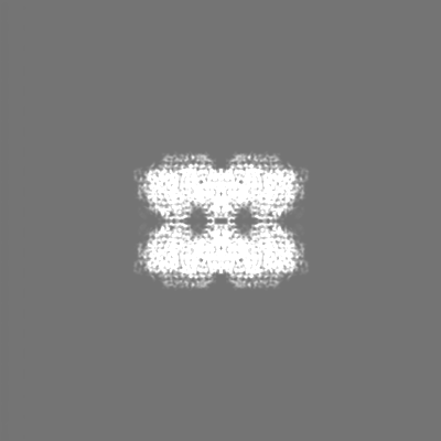
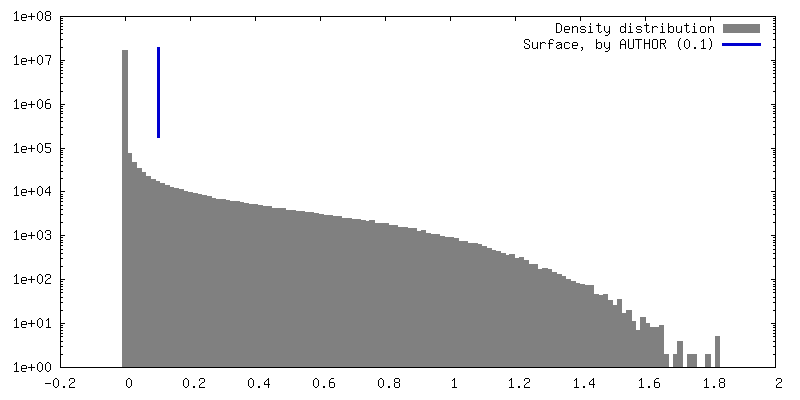
 Map data
 Sample
 Keywords
 Function and homology information
 Staphylococcus aureus (bacteria)
 Authors
 United States, 1 items
  Citation
 Structure visualization
 Downloads & links
 emd_72476.png
 http://ftp.pdbj.org/pub/emdb/structures/EMD-72476


 Z (Sec.)
 Y (Row.)
 X (Col.)


 Sample components
 Processing
 Electron microscopy
 FIELD EMISSION GUN

