Movie
Movie Controller

[English] 日本語
 Yorodumi
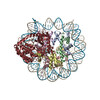
- PDB-9y46: Human nucleosome structure on Nickel-NTA lipid affinity grid (C2 ... -
+ Open data

- Basic information
| Entry | Database: PDB / ID: 9y46 | ||||||||||||
|---|---|---|---|---|---|---|---|---|---|---|---|---|---|
| Title | Human nucleosome structure on Nickel-NTA lipid affinity grid (C2 refinement) | ||||||||||||
 Components Components |
| ||||||||||||
 Keywords Keywords | DNA BINDING PROTEIN / Nucleosome / chromatin / DNA | ||||||||||||
| Function / homology |  Function and homology information Function and homology informationnegative regulation of megakaryocyte differentiation / protein localization to CENP-A containing chromatin / Chromatin modifying enzymes / nucleosomal DNA binding / Replacement of protamines by nucleosomes in the male pronucleus / CENP-A containing nucleosome / Packaging Of Telomere Ends / Regulation of PD-L1(CD274) transcription / Recognition and association of DNA glycosylase with site containing an affected purine / Cleavage of the damaged purine ...negative regulation of megakaryocyte differentiation / protein localization to CENP-A containing chromatin / Chromatin modifying enzymes / nucleosomal DNA binding / Replacement of protamines by nucleosomes in the male pronucleus / CENP-A containing nucleosome / Packaging Of Telomere Ends / Regulation of PD-L1(CD274) transcription / Recognition and association of DNA glycosylase with site containing an affected purine / Cleavage of the damaged purine / Deposition of new CENPA-containing nucleosomes at the centromere / telomere organization / Interleukin-7 signaling / Recognition and association of DNA glycosylase with site containing an affected pyrimidine / Cleavage of the damaged pyrimidine / RNA Polymerase I Promoter Opening / Inhibition of DNA recombination at telomere / Assembly of the ORC complex at the origin of replication / Meiotic synapsis / SUMOylation of chromatin organization proteins / Dengue Virus-Host Interactions / Regulation of endogenous retroelements by the Human Silencing Hub (HUSH) complex / DNA methylation / Condensation of Prophase Chromosomes / Chromatin modifications during the maternal to zygotic transition (MZT) / HCMV Late Events / SIRT1 negatively regulates rRNA expression / ERCC6 (CSB) and EHMT2 (G9a) positively regulate rRNA expression / PRC2 methylates histones and DNA / Regulation of endogenous retroelements by KRAB-ZFP proteins / Defective pyroptosis / HDACs deacetylate histones / innate immune response in mucosa / Regulation of endogenous retroelements by Piwi-interacting RNAs (piRNAs) / RNA Polymerase I Promoter Escape / Nonhomologous End-Joining (NHEJ) / Transcriptional regulation by small RNAs / HDMs demethylate histones / Formation of the beta-catenin:TCF transactivating complex / Activated PKN1 stimulates transcription of AR (androgen receptor) regulated genes KLK2 and KLK3 / RUNX1 regulates genes involved in megakaryocyte differentiation and platelet function / Negative Regulation of CDH1 Gene Transcription / NoRC negatively regulates rRNA expression / G2/M DNA damage checkpoint / PKMTs methylate histone lysines / B-WICH complex positively regulates rRNA expression / DNA Damage/Telomere Stress Induced Senescence / Meiotic recombination / Pre-NOTCH Transcription and Translation / Activation of anterior HOX genes in hindbrain development during early embryogenesis / Transcriptional regulation of granulopoiesis / RMTs methylate histone arginines / Metalloprotease DUBs / HCMV Early Events / structural constituent of chromatin / nucleosome / UCH proteinases / nucleosome assembly / antimicrobial humoral immune response mediated by antimicrobial peptide / HATs acetylate histones / E3 ubiquitin ligases ubiquitinate target proteins / Recruitment and ATM-mediated phosphorylation of repair and signaling proteins at DNA double strand breaks / antibacterial humoral response / Factors involved in megakaryocyte development and platelet production / MLL4 and MLL3 complexes regulate expression of PPARG target genes in adipogenesis and hepatic steatosis / chromatin organization / RUNX1 regulates transcription of genes involved in differentiation of HSCs / heterochromatin formation / Processing of DNA double-strand break ends / Senescence-Associated Secretory Phenotype (SASP) / Oxidative Stress Induced Senescence / Estrogen-dependent gene expression / chromosome, telomeric region / defense response to Gram-positive bacterium / Ub-specific processing proteases / Amyloid fiber formation / protein heterodimerization activity / enzyme binding / protein-containing complex / : / DNA binding / RNA binding / extracellular exosome / extracellular region / nucleoplasm / membrane / identical protein binding / nucleus Similarity search - Function | ||||||||||||
| Biological species |  Homo sapiens (human) Homo sapiens (human) | ||||||||||||
| Method | ELECTRON MICROSCOPY / single particle reconstruction / cryo EM / Resolution: 2.59 Å | ||||||||||||
 Authors Authors | Baker, R.W. / Strauss, J.D. / McGinty, R.K. / Skrajna, A. | ||||||||||||
| Funding support |  United States, 3items United States, 3items
| ||||||||||||
 Citation Citation | Journal: J Struct Biol / Year: 2025 Title: Nickel-NTA lipid-monolayer affinity grids allow for high-resolution structure determination by cryo-EM. Authors: Aleksandra Skrajna / Clara Lenger / Emily Robinson / Kevin Cannon / Reta Sarsam / Richard G Ouellette / Alberta M Abotsi / Patrick Brennwald / Robert K McGinty / Joshua D Strauss / Richard W Baker / Abstract: Grid preparation is a rate-limiting step in determining high-resolution structures by single particle cryo-EM. Particle interaction with the air-water interface often leads to denaturation, ...Grid preparation is a rate-limiting step in determining high-resolution structures by single particle cryo-EM. Particle interaction with the air-water interface often leads to denaturation, aggregation, or a preferred orientation within the ice. Some samples yield insufficient quantities of particles when using traditional grid making techniques and require the use of solid supports that concentrate samples onto the grid. Recent advances in grid-preparation show that affinity grids are promising tools to selectively concentrate proteins while simultaneously protecting samples from the air-water interface. One such technique utilizes lipid monolayers containing a lipid species with an affinity handle. Some of the first affinity grids used a holey carbon layer coated with nickel nitrilotriacetic acid (Ni-NTA) lipid, which allowed for the binding of proteins bearing the commonly used poly-histidine affinity tag. These studies however used complicated protocols and were conducted before the "resolution revolution" of cryo-EM. Here, we provide a straightforward preparation method and systematic analysis of Ni-NTA lipid monolayers as a tool for high-resolution single particle cryo-EM. We found the lipid affinity grids concentrate particles away from the AWI in thin ice (∼30 nm). We determined three structures ranging from 2.4 to 3.0 Å resolution, showing this method is amenable to high-resolution. Furthermore, we determined a 3.1 Å structure of a sub-100 kDa protein without symmetry, demonstrating the utility for a range of biological macromolecules. Lipid monolayers are therefore an easily extendable tool for most systems and help alleviate common problems such as low yield, disruption by the air-water interface, and thicker ice. | ||||||||||||
| History |
|
- Structure visualization
| Structure viewer | Molecule: MolmilJmol/JSmol |
|---|
- Downloads & links
-Download
| PDBx/mmCIF format | 9y46.cif.gz | 319.7 KB | Display | PDBx/mmCIF format |
|---|---|---|---|---|
| PDB format | pdb9y46.ent.gz | 240.8 KB | Display | PDB format |
| PDBx/mmJSON format | 9y46.json.gz | Tree view | PDBx/mmJSON format | |
| Others |  Other downloads Other downloads |
-Validation report
| Arichive directory | https://data.pdbj.org/pub/pdb/validation_reports/y4/9y46ftp://data.pdbj.org/pub/pdb/validation_reports/y4/9y46 | HTTPS FTP |
|---|
-Related structure data
| Related structure data |  72472MC  9y45C  9y47C  9y48C  9y4aC M: map data used to model this data C: citing same article ( |
|---|---|
| Similar structure data |


-Links
 PDBj
PDBj


- Assembly
| Deposited unit | 
|
|---|---|
| 1 |
|
-Components
-Protein , 4 types, 8 molecules AEBFCGDH
| #1: Protein | Mass: 15289.904 Da / Num. of mol.: 2 Source method: isolated from a genetically manipulated source Source: (gene. exp.) Homo sapiens (human)Gene: H3C15, HIST2H3A, H3C14, H3F2, H3FM, HIST2H3C, H3C13, HIST2H3D Production host:  #2: Protein | Mass: 11263.231 Da / Num. of mol.: 2 Source method: isolated from a genetically manipulated source Source: (gene. exp.) Homo sapiens (human)Gene: H4C1, H4/A, H4FA, HIST1H4A, H4C2, H4/I, H4FI, HIST1H4B, H4C3, H4/G, H4FG, HIST1H4C, H4C4, H4/B, H4FB, HIST1H4D, H4C5, H4/J, H4FJ, HIST1H4E, H4C6, H4/C, H4FC, HIST1H4F, H4C8, H4/H, H4FH, ...Gene: H4C1, H4/A, H4FA, HIST1H4A, H4C2, H4/I, H4FI, HIST1H4B, H4C3, H4/G, H4FG, HIST1H4C, H4C4, H4/B, H4FB, HIST1H4D, H4C5, H4/J, H4FJ, HIST1H4E, H4C6, H4/C, H4FC, HIST1H4F, H4C8, H4/H, H4FH, HIST1H4H, H4C9, H4/M, H4FM, HIST1H4I, H4C11, H4/E, H4FE, HIST1H4J, H4C12, H4/D, H4FD, HIST1H4K, H4C13, H4/K, H4FK, HIST1H4L, H4C14, H4/N, H4F2, H4FN, HIST2H4, HIST2H4A, H4C15, H4/O, H4FO, HIST2H4B, H4-16, HIST4H4 Production host: #3: Protein | Mass: 17559.910 Da / Num. of mol.: 2 Source method: isolated from a genetically manipulated source Details: human H2A.D with an N-terminal FLAG and HIS tag / Source: (gene. exp.) Homo sapiens (human)Gene: H2AC11, H2AFP, HIST1H2AG, H2AC13, H2AFC, HIST1H2AI, H2AC15, H2AFD, HIST1H2AK, H2AC16, H2AFI, HIST1H2AL, H2AC17, H2AFN, HIST1H2AM Production host: #4: Protein | Mass: 13806.018 Da / Num. of mol.: 2 Source method: isolated from a genetically manipulated source Source: (gene. exp.) Homo sapiens (human)Gene: H2BC4, H2BFL, HIST1H2BC, H2BC6, H2BFH, HIST1H2BE, H2BC7, H2BFG, HIST1H2BF, H2BC8, H2BFA, HIST1H2BG, H2BC10, H2BFK, HIST1H2BI Production host: |
|---|
-Nucleosomal DNA - 185 ... , 2 types, 2 molecules IJ
| #5: DNA chain | Mass: 57118.363 Da / Num. of mol.: 1 / Source method: obtained synthetically / Source: (synth.) Homo sapiens (human) |
|---|---|
| #6: DNA chain | Mass: 56495.992 Da / Num. of mol.: 1 / Source method: obtained synthetically / Source: (synth.) Homo sapiens (human) |
-Details
| Has protein modification | N |
|---|
-Experimental details
-Experiment
| Experiment | Method: ELECTRON MICROSCOPY |
|---|---|
| EM experiment | Aggregation state: PARTICLE / 3D reconstruction method: single particle reconstruction |
- Sample preparation
| Component | Name: Human nucleosome core particle / Type: COMPLEX Details: Human nucleosome core particle [Flag-His-tagged H2A (hH2A.D), H2B (hH2B.C), H3 (hH3.2), and H4 (hH4)] with a 185 bp 601 nucleosome positioning DNA sequence. Entity ID: all / Source: RECOMBINANT |
|---|---|
| Molecular weight | Experimental value: NO |
| Source (natural) | Organism: Homo sapiens (human) |
| Source (recombinant) | Organism: |
| Buffer solution | pH: 7.5 |
| Specimen | Conc.: 0.05 mg/ml / Embedding applied: NO / Shadowing applied: NO / Staining applied: NO / Vitrification applied: YES Details: Human nucleosome core particle [Flag-His-tagged H2A (hH2A.D), H2B (hH2B.C), H3 (hH3.2), and H4 (hH4)] with a 185 bp 601 nucleosome positioning DNA sequence |
| Specimen support | Grid type: Quantifoil |
| Vitrification | Cryogen name: ETHANE-PROPANE |
- Electron microscopy imaging
| Microscopy | Model: TFS TALOS Details: Beam tilt compensation enabled and calibrated using SerialEM |
|---|---|
| Electron gun | Electron source:  FIELD EMISSION GUN / Accelerating voltage: 200 kV / Illumination mode: FLOOD BEAM FIELD EMISSION GUN / Accelerating voltage: 200 kV / Illumination mode: FLOOD BEAM |
| Electron lens | Mode: BRIGHT FIELD / Nominal magnification: 45000 X / Nominal defocus max: 2500 nm / Nominal defocus min: 500 nm / Cs: 2.7 mm / C2 aperture diameter: 70 µm / Alignment procedure: COMA FREE |
| Specimen holder | Cryogen: NITROGEN |
| Image recording | Electron dose: 60 e/Å2 / Film or detector model: GATAN K3 (6k x 4k) |
- Processing
| EM software |
| ||||||||||||||||||||||||||||||||||||||||
|---|---|---|---|---|---|---|---|---|---|---|---|---|---|---|---|---|---|---|---|---|---|---|---|---|---|---|---|---|---|---|---|---|---|---|---|---|---|---|---|---|---|
| Image processing | Details: Counting mode | ||||||||||||||||||||||||||||||||||||||||
| CTF correction | Type: PHASE FLIPPING AND AMPLITUDE CORRECTION | ||||||||||||||||||||||||||||||||||||||||
| Symmetry | Point symmetry: C2 (2 fold cyclic) | ||||||||||||||||||||||||||||||||||||||||
| 3D reconstruction | Resolution: 2.59 Å / Resolution method: FSC 0.143 CUT-OFF / Num. of particles: 182456 / Symmetry type: POINT | ||||||||||||||||||||||||||||||||||||||||
| Atomic model building | Protocol: OTHER / Space: REAL | ||||||||||||||||||||||||||||||||||||||||
| Atomic model building | PDB-ID: 7JO9 Accession code: 7JO9 / Source name: PDB / Type: experimental model | ||||||||||||||||||||||||||||||||||||||||
| Refinement | Highest resolution: 2.59 Å Stereochemistry target values: REAL-SPACE (WEIGHTED MAP SUM AT ATOM CENTERS) |