 ムービー
ムービー コントローラー


+ データを開く

- 基本情報
基本情報
| 登録情報 | データベース: EMDB / ID: EMD-5410 | |||||||||
|---|---|---|---|---|---|---|---|---|---|---|
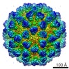
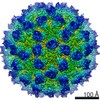
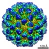
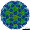
| タイトル | Cryo-electron microscopic reconstruction of wild rabbit hemorrhagic disease virus | |||||||||
 マップデータ マップデータ | Reconstruction of wild rabbit hemorrhagic disease virus; The final map was sharpened using b factor -300 A2 to 4.5 angstrom and low passed to 5 angstrom. | |||||||||
 試料 試料 |
| |||||||||
 キーワード キーワード | calicivirus / Lagovirus / rabbit hemorrhagic disease | |||||||||
| 機能・相同性 |  機能・相同性情報 機能・相同性情報calicivirin / RNA-protein covalent cross-linking / nucleoside-triphosphate phosphatase / host cell cytoplasm / membrane => GO:0016020 / RNA helicase activity / RNA-directed RNA polymerase / viral RNA genome replication / cysteine-type endopeptidase activity / RNA-dependent RNA polymerase activity ...calicivirin / RNA-protein covalent cross-linking / nucleoside-triphosphate phosphatase / host cell cytoplasm / membrane => GO:0016020 / RNA helicase activity / RNA-directed RNA polymerase / viral RNA genome replication / cysteine-type endopeptidase activity / RNA-dependent RNA polymerase activity / DNA-templated transcription / RNA binding / ATP binding / cytoplasm 類似検索 - 分子機能 | |||||||||
| 生物種 |  Rabbit hemorrhagic disease virus (ウサギ出血病ウイルス) Rabbit hemorrhagic disease virus (ウサギ出血病ウイルス) | |||||||||
| 手法 | 単粒子再構成法 / クライオ電子顕微鏡法 / 解像度: 6.5 Å | |||||||||
 データ登録者 データ登録者 | Wang X / Xu F / Liu J / Gao B / Zhang K / Zhai Y / Ma J / Hu Z / Pang X / Zheng D ...Wang X / Xu F / Liu J / Gao B / Zhang K / Zhai Y / Ma J / Hu Z / Pang X / Zheng D / Pang H / Sun F | |||||||||
 引用 引用 | ジャーナル: PLoS Pathog / 年: 2013 タイトル: Atomic model of rabbit hemorrhagic disease virus by cryo-electron microscopy and crystallography. 著者: Xue Wang / Fengting Xu / Jiasen Liu / Bingquan Gao / Yanxin Liu / Yujia Zhai / Jun Ma / Kai Zhang / Timothy S Baker / Klaus Schulten / Dong Zheng / Hai Pang / Fei Sun /  要旨: Rabbit hemorrhagic disease, first described in China in 1984, causes hemorrhagic necrosis of the liver. Its etiological agent, rabbit hemorrhagic disease virus (RHDV), belongs to the Lagovirus genus ...Rabbit hemorrhagic disease, first described in China in 1984, causes hemorrhagic necrosis of the liver. Its etiological agent, rabbit hemorrhagic disease virus (RHDV), belongs to the Lagovirus genus in the family Caliciviridae. The detailed molecular structure of any lagovirus capsid has yet to be determined. Here, we report a cryo-electron microscopic (cryoEM) reconstruction of wild-type RHDV at 6.5 Å resolution and the crystal structures of the shell (S) and protruding (P) domains of its major capsid protein, VP60, each at 2.0 Å resolution. From these data we built a complete atomic model of the RHDV capsid. VP60 has a conserved S domain and a specific P2 sub-domain that differs from those found in other caliciviruses. As seen in the shell portion of the RHDV cryoEM map, which was resolved to ~5.5 Å, the N-terminal arm domain of VP60 folds back onto its cognate S domain. Sequence alignments of VP60 from six groups of RHDV isolates revealed seven regions of high variation that could be mapped onto the surface of the P2 sub-domain and suggested three putative pockets might be responsible for binding to histo-blood group antigens. A flexible loop in one of these regions was shown to interact with rabbit tissue cells and contains an important epitope for anti-RHDV antibody production. Our study provides a reliable, pseudo-atomic model of a Lagovirus and suggests a new candidate for an efficient vaccine that can be used to protect rabbits from RHDV infection. | |||||||||
| 履歴 |
|
- 構造の表示
構造の表示
| ムービー |
ムービービューア |
|---|---|
| 構造ビューア | EMマップ: SurfViewMolmilJmol/JSmol |
| 添付画像 |

- ダウンロードとリンク
-EMDBアーカイブ
| マップデータ | emd_5410.map.gz | 326.3 MB | EMDBマップデータ形式 | |
|---|---|---|---|---|
| ヘッダ (付随情報) | emd-5410-v30.xmlemd-5410.xml | 13.1 KB 13.1 KB | 表示 表示 | EMDBヘッダ |
| 画像 | 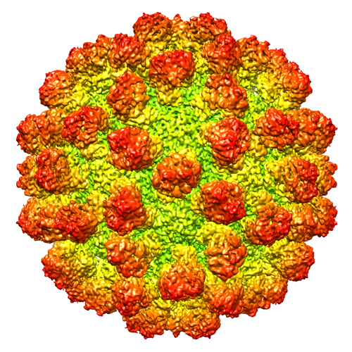 emd_5410.jpg emd_5410.jpg | 390 KB | ||
| アーカイブディレクトリ |  http://ftp.pdbj.org/pub/emdb/structures/EMD-5410ftp://ftp.pdbj.org/pub/emdb/structures/EMD-5410 http://ftp.pdbj.org/pub/emdb/structures/EMD-5410ftp://ftp.pdbj.org/pub/emdb/structures/EMD-5410 | HTTPS FTP |
-検証レポート
| 文書・要旨 | emd_5410_validation.pdf.gz | 188.5 KB | 表示 | EMDB検証レポート |
|---|---|---|---|---|
| 文書・詳細版 | emd_5410_full_validation.pdf.gz | 188.1 KB | 表示 | |
| XML形式データ | emd_5410_validation.xml.gz | 499 B | 表示 | |
| アーカイブディレクトリ | https://ftp.pdbj.org/pub/emdb/validation_reports/EMD-5410ftp://ftp.pdbj.org/pub/emdb/validation_reports/EMD-5410 | HTTPS FTP |
-関連構造データ


-リンク
| EMDBのページ | EMDB (EBI/PDBe) / EMDataResource |
|---|---|
| 「今月の分子」の関連する項目 |


-マップ
| ファイル | ダウンロード / ファイル: emd_5410.map.gz / 形式: CCP4 / 大きさ: 412 MB / タイプ: IMAGE STORED AS FLOATING POINT NUMBER (4 BYTES) | ||||||||||||||||||||||||||||||||||||||||||||||||||||||||||||||||||||
|---|---|---|---|---|---|---|---|---|---|---|---|---|---|---|---|---|---|---|---|---|---|---|---|---|---|---|---|---|---|---|---|---|---|---|---|---|---|---|---|---|---|---|---|---|---|---|---|---|---|---|---|---|---|---|---|---|---|---|---|---|---|---|---|---|---|---|---|---|---|
| 注釈 | Reconstruction of wild rabbit hemorrhagic disease virus; The final map was sharpened using b factor -300 A2 to 4.5 angstrom and low passed to 5 angstrom. | ||||||||||||||||||||||||||||||||||||||||||||||||||||||||||||||||||||
| ボクセルのサイズ | X=Y=Z: 0.933 Å | ||||||||||||||||||||||||||||||||||||||||||||||||||||||||||||||||||||
| 密度 |
| ||||||||||||||||||||||||||||||||||||||||||||||||||||||||||||||||||||
| 対称性 | 空間群: 1 | ||||||||||||||||||||||||||||||||||||||||||||||||||||||||||||||||||||
| 詳細 | EMDB XML:
CCP4マップ ヘッダ情報:
| ||||||||||||||||||||||||||||||||||||||||||||||||||||||||||||||||||||
-添付データ
- 試料の構成要素
-全体 : Wild Rabbit Hemorrhagic Disease Virus (strain HYD)
| 全体 | 名称: Wild Rabbit Hemorrhagic Disease Virus (strain HYD) |
|---|---|
| 要素 |
|
-超分子 #1000: Wild Rabbit Hemorrhagic Disease Virus (strain HYD)
| 超分子 | 名称: Wild Rabbit Hemorrhagic Disease Virus (strain HYD) / タイプ: sample / ID: 1000 / Number unique components: 1 |
|---|---|
| 分子量 | 理論値: 10.8 MDa |
-超分子 #1: Rabbit hemorrhagic disease virus
| 超分子 | 名称: Rabbit hemorrhagic disease virus / タイプ: virus / ID: 1 / Name.synonym: RHDV / 詳細: HYD strain and gene bank number is AEB26305. / NCBI-ID: 11976 / 生物種: Rabbit hemorrhagic disease virus / データベース: NCBI / ウイルスタイプ: VIRION / ウイルス・単離状態: STRAIN / ウイルス・エンベロープ: No / ウイルス・中空状態: No / Syn species name: RHDV |
|---|---|
| 宿主 | 生物種:  |
| 分子量 | 理論値: 10.8 MDa |
| ウイルス殻 | Shell ID: 1 / 名称: VP60 / 直径: 440 Å / T番号(三角分割数): 3 |
-実験情報
-構造解析
| 手法 | クライオ電子顕微鏡法 |
|---|---|
 解析 解析 | 単粒子再構成法 |
| 試料の集合状態 | particle |
-試料調製
| 緩衝液 | pH: 7 / 詳細: 50mM Tris, 50mM NaCl, 5mM EDTA |
|---|---|
| グリッド | 詳細: 200 mesh copper grid with holey array carbon support (GiG), glow discharged |
| 凍結 | 凍結剤: ETHANE / チャンバー内湿度: 100 % / チャンバー内温度: 100 K / 装置: FEI VITROBOT MARK IV / 手法: Blot for 3 seconds before plunging |
- 電子顕微鏡法
| 顕微鏡 | FEI TITAN KRIOS |
|---|---|
| 温度 | 平均: 85 K |
| アライメント法 | Legacy - 非点収差: Objective lens astigmatism was corrected at 100,000 times magnification Legacy - Electron beam tilt params: 0 |
| 日付 | 2011年1月22日 |
| 撮影 | カテゴリ: CCD フィルム・検出器のモデル: GATAN ULTRASCAN 4000 (4k x 4k) デジタル化 - サンプリング間隔: 15 µm / 実像数: 1100 / 平均電子線量: 20 e/Å2 / ビット/ピクセル: 32 |
| Tilt angle min | 0 |
| Tilt angle max | 0 |
| 電子線 | 加速電圧: 300 kV / 電子線源:  FIELD EMISSION GUN FIELD EMISSION GUN |
| 電子光学系 | 倍率(補正後): 160770 / 照射モード: FLOOD BEAM / 撮影モード: BRIGHT FIELD / Cs: 2.7 mm / 最大 デフォーカス(公称値): 2.5 µm / 最小 デフォーカス(公称値): 1.5 µm / 倍率(公称値): 96000 |
| 試料ステージ | 試料ホルダー: Liquid nitrogen cooled 試料ホルダーモデル: FEI TITAN KRIOS AUTOGRID HOLDER |
| 実験機器 |  モデル: Titan Krios / 画像提供: FEI Company |
-画像解析
| 詳細 | The particles were selected using an automatic selection program FindEM. |
|---|---|
| CTF補正 | 詳細: CTF correction of each whole micrograph |
| 最終 再構成 | アルゴリズム: OTHER / 解像度のタイプ: BY AUTHOR / 解像度: 6.5 Å / 解像度の算出法: FSC 0.5 CUT-OFF / ソフトウェア - 名称: EMAN1.9, Spider 詳細: The final map was sharpened to 4.5 Angstrom with B factor -300 and then filtered to 5.0 Angstrom. 使用した粒子像数: 26000 |
| 最終 2次元分類 | クラス数: 475 |
-原子モデル構築 1
| 初期モデル | PDB ID: Chain - Chain ID: A |
|---|---|
| ソフトウェア | 名称: Chimera |
| 詳細 | Protocol: Rigid body. The P domains were separately fitted into the map by automatic rigid body fitting using Chimera (FIT IN MAP). |
| 精密化 | 空間: REAL / プロトコル: RIGID BODY FIT |
| 得られたモデル |  PDB-3j1p: |
