- EMDB-5335: Sub-nanometer resolution structure of the intact T. thermophilus ... -
+
データを開く
IDまたはキーワード:
読み込み中...
-
基本情報
登録情報
データベース: EMDB / ID: EMD-5335
タイトル
Sub-nanometer resolution structure of the intact T. thermophilus proton-driven ATP synthase reveals the arrangement of its trans-membrane helices
マップデータ
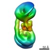
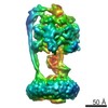
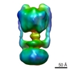
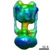
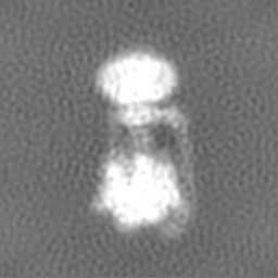
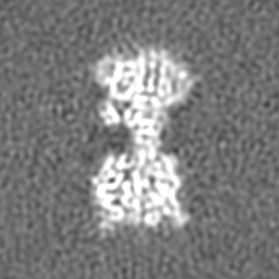
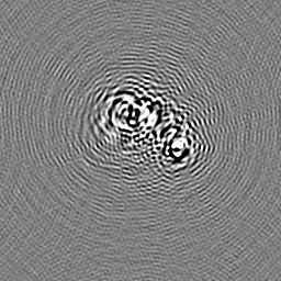
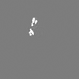
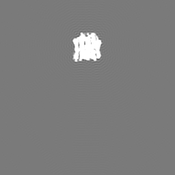
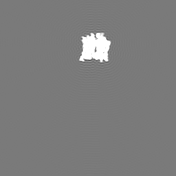
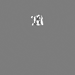
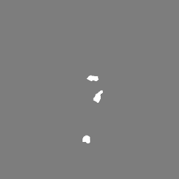
Experimental map of the V-type ATPase from Thermus thermophilus. Masks included are (a) density from subunit D not represented by the available crystal structure, (b) experimental map segment from subunit I, and (c) experimental map segment from L12-ring.
試料
試料: Thermus thermophilus V-type ATPase/ATP synthase solubilized with detergent
タンパク質・ペプチド: V-type ATPase
キーワード
ATP synthase / membrane protein complex / subnanometer / Thermus thermophilus / ATPase / V-ATPase
機能・相同性
機能・相同性情報
proton-transporting V-type ATPase, V0 domain / proton-transporting two-sector ATPase complex, catalytic domain / proton-transporting ATP synthase complex / proton motive force-driven plasma membrane ATP synthesis / H+-transporting two-sector ATPase / proton-transporting ATPase activity, rotational mechanism / proton-transporting ATP synthase activity, rotational mechanism / ATP binding / metal ion binding 類似検索 - 分子機能
ATPase, V0 complex, c subunit / : / Vacuolar (H+)-ATPase G subunit / ATPase, V1 complex, subunit F, bacterial/archaeal / ATPase, V0 complex, c/d subunit / V-type ATPase subunit C/d / V-type ATP synthase subunit c/d subunit superfamily / V-type ATP synthase c/d subunit, domain 3 superfamily / ATP synthase (C/AC39) subunit / V-type ATPase subunit E ...ATPase, V0 complex, c subunit / : / Vacuolar (H+)-ATPase G subunit / ATPase, V1 complex, subunit F, bacterial/archaeal / ATPase, V0 complex, c/d subunit / V-type ATPase subunit C/d / V-type ATP synthase subunit c/d subunit superfamily / V-type ATP synthase c/d subunit, domain 3 superfamily / ATP synthase (C/AC39) subunit / V-type ATPase subunit E / V-type ATPase subunit E, C-terminal domain superfamily / ATP synthase (E/31 kDa) subunit / ATPase, V1 complex, subunit D / ATPase, V1 complex, subunit F / ATPase, V1 complex, subunit F superfamily / ATP synthase subunit D / ATP synthase (F/14-kDa) subunit / V-type ATP synthase regulatory subunit B/beta / V-type ATP synthase catalytic alpha chain / ATPsynthase alpha/beta subunit, N-terminal extension / ATPsynthase alpha/beta subunit N-term extension / C-terminal domain of V and A type ATP synthase / ATPase, F1/V1 complex, beta/alpha subunit, C-terminal / ATP synthase subunit alpha, N-terminal domain-like superfamily / ATPase, F1/V1/A1 complex, alpha/beta subunit, N-terminal domain superfamily / ATPase, F1/V1/A1 complex, alpha/beta subunit, N-terminal domain / ATP synthase alpha/beta family, beta-barrel domain / ATPase, alpha/beta subunit, nucleotide-binding domain, active site / ATP synthase alpha and beta subunits signature. / ATPase, F1/V1/A1 complex, alpha/beta subunit, nucleotide-binding domain / ATP synthase alpha/beta family, nucleotide-binding domain / P-loop containing nucleoside triphosphate hydrolase 類似検索 - ドメイン・相同性
V-type ATP synthase subunit D / V-type ATP synthase subunit E / V-type ATP synthase subunit C / V-type ATP synthase subunit F / V-type ATP synthase alpha chain / V-type ATP synthase beta chain / V-type ATP synthase, subunit (VAPC-THERM) 類似検索 - 構成要素
ジャーナル: Nature / 年: 2011 タイトル: Subnanometre-resolution structure of the intact Thermus thermophilus H+-driven ATP synthase. 著者: Wilson C Y Lau / John L Rubinstein / 要旨: Ion-translocating rotary ATPases serve either as ATP synthases, using energy from a transmembrane ion motive force to create the cell's supply of ATP, or as transmembrane ion pumps that are powered ...Ion-translocating rotary ATPases serve either as ATP synthases, using energy from a transmembrane ion motive force to create the cell's supply of ATP, or as transmembrane ion pumps that are powered by ATP hydrolysis. The members of this family of enzymes each contain two rotary motors: one that couples ion translocation to rotation and one that couples rotation to ATP synthesis or hydrolysis. During ATP synthesis, ion translocation through the membrane-bound region of the complex causes rotation of a central rotor that drives conformational changes and ATP synthesis in the catalytic region of the complex. There are no structural models available for the intact membrane region of any ion-translocating rotary ATPase. Here we present a 9.7 Å resolution map of the H(+)-driven ATP synthase from Thermus thermophilus obtained by electron cryomicroscopy of single particles in ice. The 600-kilodalton complex has an overall subunit composition of A(3)B(3)CDE(2)FG(2)IL(12). The membrane-bound motor consists of a ring of L subunits and the carboxy-terminal region of subunit I, which are equivalent to the c and a subunits of most other rotary ATPases, respectively. The map shows that the ring contains 12 L subunits and that the I subunit has eight transmembrane helices. The L(12) ring and I subunit have a surprisingly small contact area in the middle of the membrane, with helices from the I subunit making contacts with two different L subunits. The transmembrane helices of subunit I form bundles that could serve as half-channels across the membrane, with the first half-channel conducting protons from the periplasm to the L(12) ring and the second half-channel conducting protons from the L(12) ring to the cytoplasm. This structure therefore suggests the mechanism by which a transmembrane proton motive force is converted to rotation in rotary ATPases.
ダウンロード / ファイル: emd_5335.map.gz / 形式: CCP4 / 大きさ: 62.5 MB / タイプ: IMAGE STORED AS FLOATING POINT NUMBER (4 BYTES)
注釈
Experimental map of the V-type ATPase from Thermus thermophilus. Masks included are (a) density from subunit D not represented by the available crystal structure, (b) experimental map segment from subunit I, and (c) experimental map segment from L12-ring.
凍結剤: ETHANE / チャンバー内湿度: 100 % / チャンバー内温度: 95 K / 装置: FEI VITROBOT MARK III / 詳細: Vitrification instrument: Vitrobot Mark 3 / 手法: Blot for 20 seconds before plunging
-
電子顕微鏡法
顕微鏡
FEI TECNAI F20
アライメント法
Legacy - 非点収差: Objective lens astigmatism corrected around 100,000x magnification
アルゴリズム: OTHER / 解像度のタイプ: BY AUTHOR / 解像度: 9.7 Å / 解像度の算出法: FSC 0.143 CUT-OFF ソフトウェア - 名称: Frealign, Refine_Fspace, Build_FSpace 詳細: Map constructed in Fourier space by Build_Fspace, sharpened with an inverse temperature factor of 750 Angstroms squared 使用した粒子像数: 46105
Chain - #0 - Chain ID: G / Chain - #1 - Chain ID: E
ソフトウェア
名称: UCSF Chimera
詳細
PDBEntryID_givenInChain. Protocol: Rigid Body. Two copies of this peripheral stalk (EG subcomplex) structure fitted into the cryo-EM map because there are two copies in the complex.
精密化
空間: REAL / プロトコル: RIGID BODY FIT / 当てはまり具合の基準: Correlation coefficient
得られたモデル
PDB-3j0j: Fitted atomic models of Thermus thermophilus V-ATPase subunits into cryo-EM map
 ムービー
ムービー コントローラー


 データを開く
 基本情報
基本情報 マップデータ
 試料
試料 キーワード
キーワード 機能・相同性情報
 Thermus thermophilus HB8 (バクテリア)
 データ登録者
 引用
引用
 構造の表示
構造の表示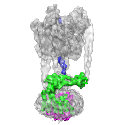
 ダウンロードとリンク
 emd_5335_1.png
 http://ftp.pdbj.org/pub/emdb/structures/EMD-5335


 Z (Sec.)
 Y (Row.)
 X (Col.)


 試料の構成要素
 解析
解析 電子顕微鏡法
 FIELD EMISSION GUN