 Movie
Movie Controller

[English] 日本語
 Yorodumi
- EMDB-52119: Translation-initiation state of human mitochondrial ribosome smal... -
+ Open data

- Basic information
| Entry |  | |||||||||||||||||||||
|---|---|---|---|---|---|---|---|---|---|---|---|---|---|---|---|---|---|---|---|---|---|---|
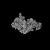
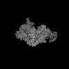
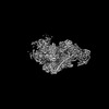
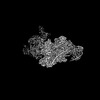
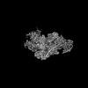
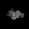
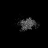
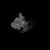
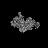
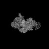
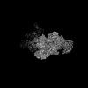
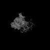
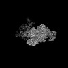
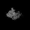
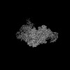
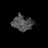
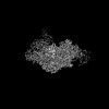
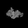
| Title | Translation-initiation state of human mitochondrial ribosome small subunit (State G) | |||||||||||||||||||||
 Map data Map data | Composite map of state G | |||||||||||||||||||||
 Sample Sample |
| |||||||||||||||||||||
 Keywords Keywords | mitochondrial ribosomal small subunit / translation-initiation intermediate / immature h44 / single-particle cryo-EM / RIBOSOME | |||||||||||||||||||||
| Function / homology |  Function and homology information Function and homology informationmitochondrial translational initiation / translation factor activity, RNA binding / formation of translation preinitiation complex / mitochondrial ribosome assembly / Mitochondrial translation elongation / Mitochondrial translation initiation / Mitochondrial ribosome-associated quality control / Mitochondrial translation termination / mitochondrial ribosome / ribosome disassembly ...mitochondrial translational initiation / translation factor activity, RNA binding / formation of translation preinitiation complex / mitochondrial ribosome assembly / Mitochondrial translation elongation / Mitochondrial translation initiation / Mitochondrial ribosome-associated quality control / Mitochondrial translation termination / mitochondrial ribosome / ribosome disassembly / mitochondrial small ribosomal subunit / mitochondrial translation / apoptotic mitochondrial changes / positive regulation of proteolysis / ribosomal small subunit binding / sperm head-tail coupling apparatus / translation initiation factor activity / Mitochondrial protein degradation / apoptotic signaling pathway / fibrillar center / cell junction / regulation of translation / small ribosomal subunit / small ribosomal subunit rRNA binding / nuclear membrane / Hydrolases; Acting on acid anhydrides; Acting on GTP to facilitate cellular and subcellular movement / tRNA binding / cell population proliferation / mitochondrial inner membrane / rRNA binding / structural constituent of ribosome / ribosome / translation / mitochondrial matrix / protein domain specific binding / mRNA binding / GTPase activity / GTP binding / nucleolus / mitochondrion / RNA binding / nucleoplasm / nucleus / plasma membrane / cytoplasm / cytosol Similarity search - Function | |||||||||||||||||||||
| Biological species |  Homo sapiens (human) Homo sapiens (human) | |||||||||||||||||||||
| Method | single particle reconstruction / cryo EM / Resolution: 3.1 Å | |||||||||||||||||||||
 Authors Authors | Finke AF / Heinrichs M / Aibara S / Richter-Dennerlein R / Hillen HS | |||||||||||||||||||||
| Funding support |  Germany, European Union, 6 items Germany, European Union, 6 items
| |||||||||||||||||||||
 Citation Citation | Journal: Nat Commun / Year: 2025 Title: Coupling of ribosome biogenesis and translation initiation in human mitochondria. Authors: Marleen Heinrichs / Anna Franziska Finke / Shintaro Aibara / Angelique Krempler / Angela Boshnakovska / Peter Rehling / Hauke S Hillen / Ricarda Richter-Dennerlein / Abstract: Biogenesis of mitoribosomes requires dedicated chaperones, RNA-modifying enzymes, and GTPases, and defects in mitoribosome assembly lead to severe mitochondriopathies in humans. Here, we characterize ...Biogenesis of mitoribosomes requires dedicated chaperones, RNA-modifying enzymes, and GTPases, and defects in mitoribosome assembly lead to severe mitochondriopathies in humans. Here, we characterize late-step assembly states of the small mitoribosomal subunit (mtSSU) by combining genetic perturbation and mutagenesis analysis with biochemical and structural approaches. Isolation of native mtSSU biogenesis intermediates via a FLAG-tagged variant of the GTPase MTG3 reveals three distinct assembly states, which show how factors cooperate to mature the 12S rRNA. In addition, we observe four distinct primed initiation mtSSU states with an incompletely matured rRNA, suggesting that biogenesis and translation initiation are not mutually exclusive processes but can occur simultaneously. Together, these results provide insights into mtSSU biogenesis and suggest a functional coupling between ribosome biogenesis and translation initiation in human mitochondria. | |||||||||||||||||||||
| History |
|
- Structure visualization
| Supplemental images |
|---|

- Downloads & links
-EMDB archive
| Map data | emd_52119.map.gz | 299.4 MB | EMDB map data format | |
|---|---|---|---|---|
| Header (meta data) | emd-52119-v30.xmlemd-52119.xml | 69.3 KB 69.3 KB | Display Display | EMDB header |
| Images |  emd_52119.png emd_52119.png | 91.3 KB | ||
| Filedesc metadata | emd-52119.cif.gz | 14.6 KB | ||
| Archive directory |  http://ftp.pdbj.org/pub/emdb/structures/EMD-52119ftp://ftp.pdbj.org/pub/emdb/structures/EMD-52119 http://ftp.pdbj.org/pub/emdb/structures/EMD-52119ftp://ftp.pdbj.org/pub/emdb/structures/EMD-52119 | HTTPS FTP |
-Related structure data
| Related structure data | 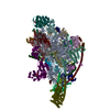 9hfoMC  9g5bC 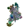 9g5cC 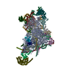 9g5dC 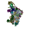 9g5eC 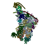 9hfmC 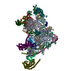 9hfnC C: citing same article ( M: atomic model generated by this map |
|---|---|
| Similar structure data |


-Links
| EMDB pages | EMDB (EBI/PDBe) / EMDataResource |
|---|---|
| Related items in Molecule of the Month |


-Map
| File | Download / File: emd_52119.map.gz / Format: CCP4 / Size: 325 MB / Type: IMAGE STORED AS FLOATING POINT NUMBER (4 BYTES) | ||||||||||||||||||||||||||||||||||||
|---|---|---|---|---|---|---|---|---|---|---|---|---|---|---|---|---|---|---|---|---|---|---|---|---|---|---|---|---|---|---|---|---|---|---|---|---|---|
| Annotation | Composite map of state G | ||||||||||||||||||||||||||||||||||||
| Projections & slices | Image control
Images are generated by Spider. | ||||||||||||||||||||||||||||||||||||
| Voxel size | X=Y=Z: 1.05 Å | ||||||||||||||||||||||||||||||||||||
| Density |
| ||||||||||||||||||||||||||||||||||||
| Symmetry | Space group: 1 | ||||||||||||||||||||||||||||||||||||
| Details | EMDB XML:
|
 Z (Sec.)
 Y (Row.)
 X (Col.)


-Supplemental data
- Sample components
+Entire : Translation-initiation state of human mitochondrial ribosome smal...
+Supramolecule #1: Translation-initiation state of human mitochondrial ribosome smal...
+Macromolecule #1: 28S ribosomal protein S34, mitochondrial
+Macromolecule #2: 28S ribosomal protein S35, mitochondrial
+Macromolecule #3: Small ribosomal subunit protein mS37
+Macromolecule #4: Aurora kinase A-interacting protein
+Macromolecule #5: Pentatricopeptide repeat domain-containing protein 3, mitochondrial
+Macromolecule #8: Translation initiation factor IF-2, mitochondrial
+Macromolecule #10: 28S ribosomal protein S2, mitochondrial
+Macromolecule #11: 28S ribosomal protein S24, mitochondrial
+Macromolecule #12: 28S ribosomal protein S5, mitochondrial
+Macromolecule #13: 28S ribosomal protein S6, mitochondrial
+Macromolecule #14: 28S ribosomal protein S7, mitochondrial
+Macromolecule #15: 28S ribosomal protein S9, mitochondrial
+Macromolecule #16: 28S ribosomal protein S10, mitochondrial
+Macromolecule #17: 28S ribosomal protein S11, mitochondrial
+Macromolecule #18: 28S ribosomal protein S12, mitochondrial
+Macromolecule #19: 28S ribosomal protein S14, mitochondrial
+Macromolecule #20: 28S ribosomal protein S15, mitochondrial
+Macromolecule #21: 28S ribosomal protein S16, mitochondrial
+Macromolecule #22: 28S ribosomal protein S17, mitochondrial
+Macromolecule #23: 28S ribosomal protein S18b, mitochondrial
+Macromolecule #24: 28S ribosomal protein S18c, mitochondrial
+Macromolecule #25: 28S ribosomal protein S21, mitochondrial
+Macromolecule #26: 28S ribosomal protein S22, mitochondrial
+Macromolecule #27: 28S ribosomal protein S23, mitochondrial
+Macromolecule #28: 28S ribosomal protein S25, mitochondrial
+Macromolecule #29: 28S ribosomal protein S26, mitochondrial
+Macromolecule #30: 28S ribosomal protein S27, mitochondrial
+Macromolecule #31: 28S ribosomal protein S28, mitochondrial
+Macromolecule #32: 28S ribosomal protein S29, mitochondrial
+Macromolecule #33: 28S ribosomal protein S31, mitochondrial
+Macromolecule #34: 28S ribosomal protein S33, mitochondrial
+Macromolecule #6: fMet-tRNAMet
+Macromolecule #7: mRNA, start codon
+Macromolecule #9: 12S mitochondrial rRNA
+Macromolecule #35: MAGNESIUM ION
+Macromolecule #36: GUANOSINE-5'-TRIPHOSPHATE

+Macromolecule #37: POTASSIUM ION
+Macromolecule #38: ZINC ION
+Macromolecule #39: FE2/S2 (INORGANIC) CLUSTER

+Macromolecule #40: ADENOSINE-5'-TRIPHOSPHATE

+Macromolecule #41: GUANOSINE-5'-DIPHOSPHATE

-Experimental details
-Structure determination
| Method | cryo EM |
|---|---|
 Processing Processing | single particle reconstruction |
| Aggregation state | particle |
-Sample preparation
| Buffer | pH: 7.4 Component:
Details: 20mM HEPES-HCl, 100mM KCl, 20mM MgCl2, 0.02% DDM, 1mM PMSF, 1x protease-inhibitor mix, 0.5mM GMP-PNP | ||||||||||||||||||||||||
|---|---|---|---|---|---|---|---|---|---|---|---|---|---|---|---|---|---|---|---|---|---|---|---|---|---|
| Grid | Model: Quantifoil R3.5/1 / Material: COPPER / Support film - #0 - Film type ID: 1 / Support film - #0 - Material: CARBON / Support film - #0 - topology: HOLEY / Support film - #1 - Film type ID: 2 / Support film - #1 - Material: CARBON / Support film - #1 - topology: CONTINUOUS / Pretreatment - Type: GLOW DISCHARGE / Details: The grid was precoated with a 2-3 nm carbon layer. | ||||||||||||||||||||||||
| Vitrification | Cryogen name: ETHANE / Chamber humidity: 95 % / Chamber temperature: 277.15 K / Instrument: FEI VITROBOT MARK IV | ||||||||||||||||||||||||
| Details | The sample was crosslinked with 0.15% glutaraldehyde for 10 min on ice. The reaction was stopped by adding 50 mM lysine pH 7.5 and 50 mM aspartate pH 7.5, and subsequently desalted against the final buffer prior to vitrification. |
- Electron microscopy
| Microscope | TFS KRIOS |
|---|---|
| Image recording | Film or detector model: GATAN K3 BIOQUANTUM (6k x 4k) / Average exposure time: 3.0 sec. / Average electron dose: 40.0 e/Å2 |
| Electron beam | Acceleration voltage: 300 kV / Electron source:  FIELD EMISSION GUN FIELD EMISSION GUN |
| Electron optics | Illumination mode: OTHER / Imaging mode: BRIGHT FIELD / Cs: 2.7 mm / Nominal defocus max: 1.6 µm / Nominal defocus min: 0.6 µm / Nominal magnification: 81000 |
| Sample stage | Specimen holder model: FEI TITAN KRIOS AUTOGRID HOLDER / Cooling holder cryogen: NITROGEN |
| Experimental equipment |  Model: Titan Krios / Image courtesy: FEI Company |
+Image processing
-Atomic model buiding 1
| Initial model |
| ||||||||||||||||||||||||||||||||||||||||||||||||||||||||||||||||||||||
|---|---|---|---|---|---|---|---|---|---|---|---|---|---|---|---|---|---|---|---|---|---|---|---|---|---|---|---|---|---|---|---|---|---|---|---|---|---|---|---|---|---|---|---|---|---|---|---|---|---|---|---|---|---|---|---|---|---|---|---|---|---|---|---|---|---|---|---|---|---|---|---|
| Refinement | Space: REAL / Protocol: RIGID BODY FIT | ||||||||||||||||||||||||||||||||||||||||||||||||||||||||||||||||||||||
| Output model | PDB-9hfo: |

