Movie
Movie Controller

[English] 日本語
 Yorodumi
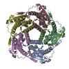
- EMDB-51006: Circularly permuted lumazine synthase 12-pentamer spherical cage -
+ Open data

- Basic information
| Entry |  | ||||||||||||
|---|---|---|---|---|---|---|---|---|---|---|---|---|---|
| Title | Circularly permuted lumazine synthase 12-pentamer spherical cage | ||||||||||||
 Map data Map data | Combined half maps post-processed by DeepEMhancer with highRes normalization mode. | ||||||||||||
 Sample Sample |
| ||||||||||||
 Keywords Keywords | protein cage / protein engineering / self-assembly / geometry / helical reconstruction / bionanotechnology / polymorphism / pentamer / DE NOVO PROTEIN | ||||||||||||
| Function / homology |  Function and homology information Function and homology information6,7-dimethyl-8-ribityllumazine synthase / 6,7-dimethyl-8-ribityllumazine synthase activity / riboflavin synthase complex / riboflavin biosynthetic process / cytoplasm / cytosol Similarity search - Function | ||||||||||||
| Biological species |   Aquifex aeolicus VF5 (bacteria) Aquifex aeolicus VF5 (bacteria) | ||||||||||||
| Method | single particle reconstruction / cryo EM / Resolution: 2.08 Å | ||||||||||||
 Authors Authors | Koziej L / Azuma Y | ||||||||||||
| Funding support |  Poland, European Union, 3 items Poland, European Union, 3 items
| ||||||||||||
 Citation Citation | Journal: ACS Nano / Year: 2025 Title: Dynamic Assembly of Pentamer-Based Protein Nanotubes. Authors: Lukasz Koziej / Farzad Fatehi / Marta Aleksejczuk / Matthew J Byrne / Jonathan G Heddle / Reidun Twarock / Yusuke Azuma /  Abstract: Hollow proteinaceous particles are useful nanometric containers for delivery and catalysis. Understanding the molecular mechanisms and the geometrical theory behind the polymorphic protein assemblies ...Hollow proteinaceous particles are useful nanometric containers for delivery and catalysis. Understanding the molecular mechanisms and the geometrical theory behind the polymorphic protein assemblies provides a basis for designing ones with the desired morphology. As such, we found that a circularly permuted variant of a cage-forming enzyme, lumazine synthase, cpAaLS, assembles into a variety of hollow spherical and cylindrical structures in response to changes in ionic strength. Cryogenic electron microscopy revealed that these structures are composed entirely of pentameric subunits, and the dramatic cage-to-tube transformation is attributed to the moderately hindered 3-fold symmetry interaction and the imparted torsion angle of the building blocks, where both mechanisms are mediated by an α-helix domain that is untethered from the native position by circular permutation. Mathematical modeling suggests that the unique double- and triple-stranded helical arrangements of subunits are optimal tiling patterns, while different geometries should be possible by modulating the interaction angles of the pentagons. These structural insights into dynamic, pentamer-based protein cages and nanotubes afford guidelines for designing nanoarchitectures with customized morphology and assembly characteristics. | ||||||||||||
| History |
|
- Structure visualization
| Supplemental images |
|---|

- Downloads & links
-EMDB archive
| Map data | emd_51006.map.gz | 70.5 MB | EMDB map data format | |
|---|---|---|---|---|
| Header (meta data) | emd-51006-v30.xmlemd-51006.xml | 26.3 KB 26.3 KB | Display Display | EMDB header |
| FSC (resolution estimation) | emd_51006_fsc.xml | 9.9 KB | Display | FSC data file |
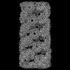
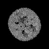
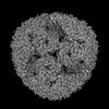
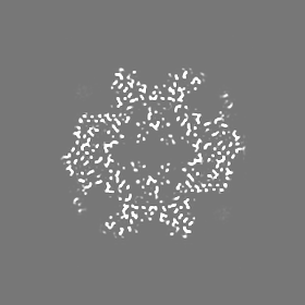
| Images |  emd_51006.png emd_51006.png | 238.6 KB | ||
| Masks | emd_51006_msk_1.map | 83.7 MB | Mask map | |
| Filedesc metadata | emd-51006.cif.gz | 7 KB | ||
| Others | emd_51006_additional_1.map.gzemd_51006_additional_2.map.gzemd_51006_half_map_1.map.gzemd_51006_half_map_2.map.gz | 64.9 MB 78.2 MB 65.3 MB 65.3 MB | ||
| Archive directory |  http://ftp.pdbj.org/pub/emdb/structures/EMD-51006ftp://ftp.pdbj.org/pub/emdb/structures/EMD-51006 http://ftp.pdbj.org/pub/emdb/structures/EMD-51006ftp://ftp.pdbj.org/pub/emdb/structures/EMD-51006 | HTTPS FTP |
-Related structure data
| Related structure data | 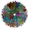 9g3pMC 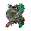 9g3hC 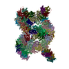 9g3iC 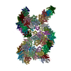 9g3jC 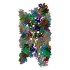 9g3mC 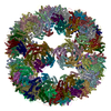 9g3nC 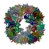 9g3oC M: atomic model generated by this map C: citing same article ( |
|---|---|
| Similar structure data |
-Links
| EMDB pages | EMDB (EBI/PDBe) / EMDataResource |
|---|
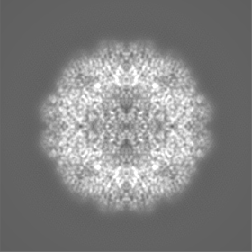
-Map
| File | Download / File: emd_51006.map.gz / Format: CCP4 / Size: 83.7 MB / Type: IMAGE STORED AS FLOATING POINT NUMBER (4 BYTES) | ||||||||||||||||||||||||||||||||||||
|---|---|---|---|---|---|---|---|---|---|---|---|---|---|---|---|---|---|---|---|---|---|---|---|---|---|---|---|---|---|---|---|---|---|---|---|---|---|
| Annotation | Combined half maps post-processed by DeepEMhancer with highRes normalization mode. | ||||||||||||||||||||||||||||||||||||
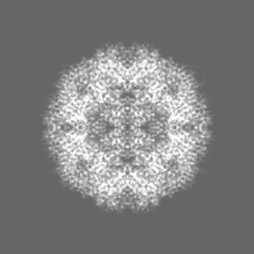
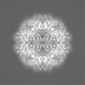
| Projections & slices | Image control
Images are generated by Spider. | ||||||||||||||||||||||||||||||||||||
| Voxel size | X=Y=Z: 0.86 Å | ||||||||||||||||||||||||||||||||||||
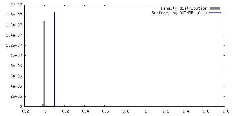
| Density |
| ||||||||||||||||||||||||||||||||||||
| Symmetry | Space group: 1 | ||||||||||||||||||||||||||||||||||||
| Details | EMDB XML:
|
 Z (Sec.)
 Y (Row.)
 X (Col.)


-Supplemental data
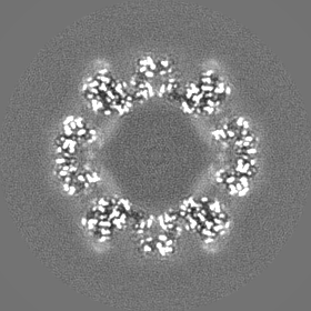
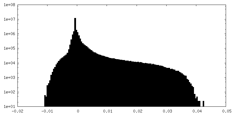
-Mask #1
| File | emd_51006_msk_1.map | ||||||||||||
|---|---|---|---|---|---|---|---|---|---|---|---|---|---|
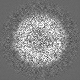
| Projections & Slices |
| ||||||||||||
| Density Histograms |


-Additional map: Map combined from 2 independent halves. Not post-processed.
| File | emd_51006_additional_1.map | ||||||||||||
|---|---|---|---|---|---|---|---|---|---|---|---|---|---|
| Annotation | Map combined from 2 independent halves. Not post-processed. | ||||||||||||
| Projections & Slices |
| ||||||||||||
| Density Histograms |


-Additional map: Combined half maps post-processed using sharpening B factor 65.0.
| File | emd_51006_additional_2.map | ||||||||||||
|---|---|---|---|---|---|---|---|---|---|---|---|---|---|
| Annotation | Combined half maps post-processed using sharpening B factor 65.0. | ||||||||||||
| Projections & Slices |
| ||||||||||||
| Density Histograms |


-Half map: #1
| File | emd_51006_half_map_1.map | ||||||||||||
|---|---|---|---|---|---|---|---|---|---|---|---|---|---|
| Projections & Slices |
| ||||||||||||
| Density Histograms |


-Half map: #2
| File | emd_51006_half_map_2.map | ||||||||||||
|---|---|---|---|---|---|---|---|---|---|---|---|---|---|
| Projections & Slices |
| ||||||||||||
| Density Histograms |


- Sample components
-Entire : Circularly permuted lumazine synthase 12-pentamer spherical cage
| Entire | Name: Circularly permuted lumazine synthase 12-pentamer spherical cage |
|---|---|
| Components |
|
-Supramolecule #1: Circularly permuted lumazine synthase 12-pentamer spherical cage
| Supramolecule | Name: Circularly permuted lumazine synthase 12-pentamer spherical cage type: complex / ID: 1 / Parent: 0 / Macromolecule list: all Details: Circularly permuted (84) variant of Aquifex aeolicus lumazine synthase |
|---|---|
| Source (natural) | Organism: Aquifex aeolicus VF5 (bacteria) |
-Macromolecule #1: 6,7-dimethyl-8-ribityllumazine synthase
| Macromolecule | Name: 6,7-dimethyl-8-ribityllumazine synthase / type: protein_or_peptide / ID: 1 / Details: cpAaLS(84),cpAaLS(84) / Number of copies: 60 / Enantiomer: LEVO / EC number: 6,7-dimethyl-8-ribityllumazine synthase |
|---|---|
| Source (natural) | Organism: Aquifex aeolicus VF5 (bacteria) |
| Molecular weight | Theoretical: 17.449938 KDa |
| Recombinant expression | Organism: |
| Sequence | String: MATPHFDYIA SEVSKGLANL SLELRKPITF GVITADTLEQ AIERAGTKHG NKGWEAALSA IEMANLFKSL RGTGGSGSSM EIYEGKLTA EGLRFGIVAS RFNHALVDRL VEGAIDCIVR HGGREEDITL VRVPGSWEIP VAAGELARKE DIDAVIAIGV L IRG UniProtKB: 6,7-dimethyl-8-ribityllumazine synthase, 6,7-dimethyl-8-ribityllumazine synthase |
-Experimental details
-Structure determination
| Method | cryo EM |
|---|---|
 Processing Processing | single particle reconstruction |
| Aggregation state | particle |
-Sample preparation
| Concentration | 1 mg/mL | |||||||||
|---|---|---|---|---|---|---|---|---|---|---|
| Buffer | pH: 8 Component:
| |||||||||
| Grid | Model: Quantifoil R2/2 / Material: COPPER / Mesh: 200 / Support film - Material: CARBON / Support film - topology: HOLEY / Pretreatment - Type: GLOW DISCHARGE / Pretreatment - Time: 60 sec. / Pretreatment - Atmosphere: AIR | |||||||||
| Vitrification | Cryogen name: ETHANE / Chamber humidity: 100 % / Chamber temperature: 283 K / Instrument: FEI VITROBOT MARK IV |
- Electron microscopy
| Microscope | TFS KRIOS |
|---|---|
| Specialist optics | Energy filter - Name: GIF Bioquantum / Energy filter - Slit width: 20 eV |
| Image recording | Film or detector model: GATAN K3 BIOCONTINUUM (6k x 4k) / Digitization - Dimensions - Width: 5760 pixel / Digitization - Dimensions - Height: 4092 pixel / Number grids imaged: 1 / Number real images: 8486 / Average electron dose: 40.0 e/Å2 |
| Electron beam | Acceleration voltage: 300 kV / Electron source:  FIELD EMISSION GUN FIELD EMISSION GUN |
| Electron optics | C2 aperture diameter: 50.0 µm / Calibrated magnification: 105000 / Illumination mode: FLOOD BEAM / Imaging mode: BRIGHT FIELD / Cs: 2.7 mm / Nominal defocus max: 2.1 µm / Nominal defocus min: 0.9 µm |
| Sample stage | Specimen holder model: FEI TITAN KRIOS AUTOGRID HOLDER / Cooling holder cryogen: NITROGEN |
| Experimental equipment |  Model: Titan Krios / Image courtesy: FEI Company |
+Image processing

-Atomic model buiding 1
| Initial model | PDB ID: Chain - Source name: PDB / Chain - Initial model type: experimental model |
|---|---|
| Details | Initial fitting was done using ChimeraX. Flexible fitting was done using Isolde. |
| Refinement | Space: REAL / Protocol: RIGID BODY FIT |
| Output model | PDB-9g3p: |