[English] 日本語
Yorodumi
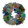
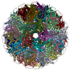
- PDB-9g3i: Circularly permuted lumazine synthase twisted tube with 18 Angstr... -

+
Open data


ID or keywords:

Loading...

-
Basic information

Entry
Database: PDB / ID: 9g3i
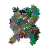
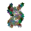
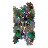
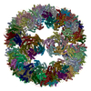
TitleCircularly permuted lumazine synthase twisted tube with 18 Angstrom gap between double strands
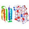
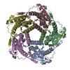
Components6,7-dimethyl-8-ribityllumazine synthase
KeywordsDE NOVO PROTEIN / protein cage / protein engineering / self-assembly / geometry / helical reconstruction / bionanotechnology / polymorphism / pentamer
Function / homology
Function and homology information


6,7-dimethyl-8-ribityllumazine synthase / 6,7-dimethyl-8-ribityllumazine synthase activity / riboflavin synthase complex / riboflavin biosynthetic process / cytosol / cytoplasm
Similarity search - Function
Lumazine synthase / Lumazine/riboflavin synthase / Lumazine/riboflavin synthase superfamily / 6,7-dimethyl-8-ribityllumazine synthase
Similarity search - Domain/homology
6,7-dimethyl-8-ribityllumazine synthase
Similarity search - Component
Biological speciesAquifex aeolicus VF5 (bacteria)
MethodELECTRON MICROSCOPY / helical reconstruction / cryo EM / Resolution: 2.85 Å
AuthorsKoziej, L. / Azuma, Y.
Funding support Poland, European Union, 3items
OrganizationGrant numberCountry
Polish National Science Centre2018/31/D/NZ1/01102 Poland
Polish National Science Centre2019/35/B/NZ1/02044 Poland
European Molecular Biology Organization (EMBO)EMBO Installation Grant (YA)European Union
CitationJournal: ACS Nano / Year: 2025
Title: Dynamic Assembly of Pentamer-Based Protein Nanotubes.
Authors: Lukasz Koziej / Farzad Fatehi / Marta Aleksejczuk / Matthew J Byrne / Jonathan G Heddle / Reidun Twarock / Yusuke Azuma /
Abstract: Hollow proteinaceous particles are useful nanometric containers for delivery and catalysis. Understanding the molecular mechanisms and the geometrical theory behind the polymorphic protein assemblies ...Hollow proteinaceous particles are useful nanometric containers for delivery and catalysis. Understanding the molecular mechanisms and the geometrical theory behind the polymorphic protein assemblies provides a basis for designing ones with the desired morphology. As such, we found that a circularly permuted variant of a cage-forming enzyme, lumazine synthase, cpAaLS, assembles into a variety of hollow spherical and cylindrical structures in response to changes in ionic strength. Cryogenic electron microscopy revealed that these structures are composed entirely of pentameric subunits, and the dramatic cage-to-tube transformation is attributed to the moderately hindered 3-fold symmetry interaction and the imparted torsion angle of the building blocks, where both mechanisms are mediated by an α-helix domain that is untethered from the native position by circular permutation. Mathematical modeling suggests that the unique double- and triple-stranded helical arrangements of subunits are optimal tiling patterns, while different geometries should be possible by modulating the interaction angles of the pentagons. These structural insights into dynamic, pentamer-based protein cages and nanotubes afford guidelines for designing nanoarchitectures with customized morphology and assembly characteristics.
History
DepositionJul 12, 2024Deposition site: PDBE / Processing site: PDBE
Revision 1.0Mar 5, 2025Provider: repository / Type: Initial release
Revision 1.0Mar 5, 2025Data content type: EM metadata / Data content type: EM metadata / Provider: repository / Type: Initial release
Revision 1.0Mar 5, 2025Data content type: Additional map / Part number: 1 / Data content type: Additional map / Provider: repository / Type: Initial release
Revision 1.0Mar 5, 2025Data content type: Additional map / Part number: 2 / Data content type: Additional map / Provider: repository / Type: Initial release
Revision 1.0Mar 5, 2025Data content type: Additional map / Part number: 3 / Data content type: Additional map / Provider: repository / Type: Initial release
Revision 1.0Mar 5, 2025Data content type: Additional map / Part number: 4 / Data content type: Additional map / Provider: repository / Type: Initial release
Revision 1.0Mar 5, 2025Data content type: FSC / Data content type: FSC / Provider: repository / Type: Initial release
Revision 1.0Mar 5, 2025Data content type: Half map / Part number: 1 / Data content type: Half map / Provider: repository / Type: Initial release
Revision 1.0Mar 5, 2025Data content type: Half map / Part number: 2 / Data content type: Half map / Provider: repository / Type: Initial release
Revision 1.0Mar 5, 2025Data content type: Image / Data content type: Image / Provider: repository / Type: Initial release
Revision 1.0Mar 5, 2025Data content type: Mask / Part number: 1 / Data content type: Mask / Provider: repository / Type: Initial release
Revision 1.0Mar 5, 2025Data content type: Mask / Part number: 2 / Data content type: Mask / Provider: repository / Type: Initial release
Revision 1.0Mar 5, 2025Data content type: Primary map / Data content type: Primary map / Provider: repository / Type: Initial release
Revision 1.1Mar 19, 2025Group: Data collection / Database references / Category: citation / em_admin
Item: _citation.journal_volume / _citation.page_first ..._citation.journal_volume / _citation.page_first / _citation.page_last / _em_admin.last_update

-
Structure visualization

Structure viewerMolecule:
MolmilJmol/JSmol

Downloads & links

-
Assembly

Deposited unit
A: 6,7-dimethyl-8-ribityllumazine synthase
B: 6,7-dimethyl-8-ribityllumazine synthase
C: 6,7-dimethyl-8-ribityllumazine synthase
D: 6,7-dimethyl-8-ribityllumazine synthase
E: 6,7-dimethyl-8-ribityllumazine synthase
F: 6,7-dimethyl-8-ribityllumazine synthase
G: 6,7-dimethyl-8-ribityllumazine synthase
H: 6,7-dimethyl-8-ribityllumazine synthase
I: 6,7-dimethyl-8-ribityllumazine synthase
J: 6,7-dimethyl-8-ribityllumazine synthase
K: 6,7-dimethyl-8-ribityllumazine synthase
L: 6,7-dimethyl-8-ribityllumazine synthase
M: 6,7-dimethyl-8-ribityllumazine synthase
N: 6,7-dimethyl-8-ribityllumazine synthase
O: 6,7-dimethyl-8-ribityllumazine synthase
P: 6,7-dimethyl-8-ribityllumazine synthase
Q: 6,7-dimethyl-8-ribityllumazine synthase
R: 6,7-dimethyl-8-ribityllumazine synthase
S: 6,7-dimethyl-8-ribityllumazine synthase
T: 6,7-dimethyl-8-ribityllumazine synthase
U: 6,7-dimethyl-8-ribityllumazine synthase
V: 6,7-dimethyl-8-ribityllumazine synthase
W: 6,7-dimethyl-8-ribityllumazine synthase
X: 6,7-dimethyl-8-ribityllumazine synthase
Y: 6,7-dimethyl-8-ribityllumazine synthase
Z: 6,7-dimethyl-8-ribityllumazine synthase
AA: 6,7-dimethyl-8-ribityllumazine synthase
BA: 6,7-dimethyl-8-ribityllumazine synthase
CA: 6,7-dimethyl-8-ribityllumazine synthase
DA: 6,7-dimethyl-8-ribityllumazine synthase
EA: 6,7-dimethyl-8-ribityllumazine synthase
FA: 6,7-dimethyl-8-ribityllumazine synthase
GA: 6,7-dimethyl-8-ribityllumazine synthase
HA: 6,7-dimethyl-8-ribityllumazine synthase
IA: 6,7-dimethyl-8-ribityllumazine synthase
JA: 6,7-dimethyl-8-ribityllumazine synthase
KA: 6,7-dimethyl-8-ribityllumazine synthase
LA: 6,7-dimethyl-8-ribityllumazine synthase
MA: 6,7-dimethyl-8-ribityllumazine synthase
NA: 6,7-dimethyl-8-ribityllumazine synthase
OA: 6,7-dimethyl-8-ribityllumazine synthase
PA: 6,7-dimethyl-8-ribityllumazine synthase
QA: 6,7-dimethyl-8-ribityllumazine synthase
RA: 6,7-dimethyl-8-ribityllumazine synthase
SA: 6,7-dimethyl-8-ribityllumazine synthase
TA: 6,7-dimethyl-8-ribityllumazine synthase
UA: 6,7-dimethyl-8-ribityllumazine synthase
VA: 6,7-dimethyl-8-ribityllumazine synthase
WA: 6,7-dimethyl-8-ribityllumazine synthase
XA: 6,7-dimethyl-8-ribityllumazine synthase
YA: 6,7-dimethyl-8-ribityllumazine synthase
ZA: 6,7-dimethyl-8-ribityllumazine synthase
AB: 6,7-dimethyl-8-ribityllumazine synthase
BB: 6,7-dimethyl-8-ribityllumazine synthase
CB: 6,7-dimethyl-8-ribityllumazine synthase
DB: 6,7-dimethyl-8-ribityllumazine synthase
EB: 6,7-dimethyl-8-ribityllumazine synthase
FB: 6,7-dimethyl-8-ribityllumazine synthase
GB: 6,7-dimethyl-8-ribityllumazine synthase
HB: 6,7-dimethyl-8-ribityllumazine synthase
IB: 6,7-dimethyl-8-ribityllumazine synthase
JB: 6,7-dimethyl-8-ribityllumazine synthase
KB: 6,7-dimethyl-8-ribityllumazine synthase
LB: 6,7-dimethyl-8-ribityllumazine synthase
MB: 6,7-dimethyl-8-ribityllumazine synthase
NB: 6,7-dimethyl-8-ribityllumazine synthase
OB: 6,7-dimethyl-8-ribityllumazine synthase
PB: 6,7-dimethyl-8-ribityllumazine synthase
QB: 6,7-dimethyl-8-ribityllumazine synthase
RB: 6,7-dimethyl-8-ribityllumazine synthase
SB: 6,7-dimethyl-8-ribityllumazine synthase
TB: 6,7-dimethyl-8-ribityllumazine synthase
UB: 6,7-dimethyl-8-ribityllumazine synthase
VB: 6,7-dimethyl-8-ribityllumazine synthase
WB: 6,7-dimethyl-8-ribityllumazine synthase
XB: 6,7-dimethyl-8-ribityllumazine synthase
YB: 6,7-dimethyl-8-ribityllumazine synthase
ZB: 6,7-dimethyl-8-ribityllumazine synthase
AC: 6,7-dimethyl-8-ribityllumazine synthase
BC: 6,7-dimethyl-8-ribityllumazine synthase
CC: 6,7-dimethyl-8-ribityllumazine synthase
DC: 6,7-dimethyl-8-ribityllumazine synthase
EC: 6,7-dimethyl-8-ribityllumazine synthase
FC: 6,7-dimethyl-8-ribityllumazine synthase
GC: 6,7-dimethyl-8-ribityllumazine synthase
HC: 6,7-dimethyl-8-ribityllumazine synthase
IC: 6,7-dimethyl-8-ribityllumazine synthase
JC: 6,7-dimethyl-8-ribityllumazine synthase
KC: 6,7-dimethyl-8-ribityllumazine synthase
LC: 6,7-dimethyl-8-ribityllumazine synthase
MC: 6,7-dimethyl-8-ribityllumazine synthase
NC: 6,7-dimethyl-8-ribityllumazine synthase
OC: 6,7-dimethyl-8-ribityllumazine synthase
PC: 6,7-dimethyl-8-ribityllumazine synthase
QC: 6,7-dimethyl-8-ribityllumazine synthase
RC: 6,7-dimethyl-8-ribityllumazine synthase
SC: 6,7-dimethyl-8-ribityllumazine synthase
TC: 6,7-dimethyl-8-ribityllumazine synthase
UC: 6,7-dimethyl-8-ribityllumazine synthase
VC: 6,7-dimethyl-8-ribityllumazine synthase


Theoretical massNumber of molelcules
Total (without water)1,746,593100
Polymers1,746,593100
Non-polymers00
Water00
1


  • Idetical with deposited unit
  • defined by author&software
  • Evidence: electron microscopy, not applicable
TypeNameSymmetry operationNumber
identity operation1_555x,y,z1

-
Components

#1: Protein ...
6,7-dimethyl-8-ribityllumazine synthase / DMRL synthase / LS / Lumazine synthase


Mass: 17465.934 Da / Num. of mol.: 100 / Mutation: 119 circular permutation,C37S,A85C
Source method: isolated from a genetically manipulated source
Details: cpAaLS(119, C37S, A85C),cpAaLS(119, C37S, A85C),cpAaLS(119, C37S, A85C),cpAaLS(119, C37S, A85C)
Source: (gene. exp.) Aquifex aeolicus VF5 (bacteria) / Gene: ribH, aq_132 / Production host: Escherichia coli #1/H766 (bacteria) / Strain (production host): BL21-Gold(DE3)
References: UniProt: O66529, 6,7-dimethyl-8-ribityllumazine synthase
Has protein modificationN

-
Experimental details

-
Experiment

ExperimentMethod: ELECTRON MICROSCOPY
EM experimentAggregation state: HELICAL ARRAY / 3D reconstruction method: helical reconstruction

-
Sample preparation

ComponentName: Circularly permuted lumazine synthase twisted tube with 18 Angstrom gap between double strands
Type: COMPLEX
Details: Circularly permuted (119) variant of Aquifex aeolicus lumazine synthase with C37S and A85C mutations
Entity ID: all / Source: RECOMBINANT
Molecular weightValue: 600 kDa/nm / Experimental value: YES
Source (natural)Organism: Aquifex aeolicus VF5 (bacteria)
Source (recombinant)Organism: Escherichia coli (E. coli) / Strain: BL21-Gold(DE3)
Buffer solutionpH: 7
Buffer component
IDConc.NameFormulaBuffer-ID
150 mMtris(hydroxymethyl)aminomethaneTris-HCl1
2150 mMsodium chlorideNaCl1
31 mMtris(2-carboxyethyl)phosphineTCEP1
SpecimenConc.: 1 mg/ml / Embedding applied: NO / Shadowing applied: NO / Staining applied: NO / Vitrification applied: YES
Specimen supportGrid material: COPPER / Grid mesh size: 200 divisions/in. / Grid type: Quantifoil R2/2
VitrificationInstrument: FEI VITROBOT MARK IV / Cryogen name: ETHANE / Humidity: 100 % / Chamber temperature: 283 K

-
Electron microscopy imaging

Experimental equipment
Model: Titan Krios / Image courtesy: FEI Company
MicroscopyModel: TFS KRIOS
Electron gunElectron source: FIELD EMISSION GUN / Accelerating voltage: 300 kV / Illumination mode: FLOOD BEAM
Electron lensMode: BRIGHT FIELD / Calibrated magnification: 105000 X / Nominal defocus max: 1500 nm / Nominal defocus min: 900 nm / Cs: 2.7 mm / C2 aperture diameter: 50 µm / Alignment procedure: COMA FREE
Specimen holderCryogen: NITROGEN / Specimen holder model: FEI TITAN KRIOS AUTOGRID HOLDER
Image recordingElectron dose: 42.44 e/Å2 / Film or detector model: GATAN K3 BIOQUANTUM (6k x 4k) / Num. of grids imaged: 1 / Num. of real images: 12746
EM imaging opticsEnergyfilter name: GIF Bioquantum / Energyfilter slit width: 20 eV
Image scansWidth: 5760 / Height: 4092

-
Processing

EM software
IDNameVersionCategoryDetails
1cryoSPARC4.4.1particle selection
2EPU2.10.0.1941RELimage acquisition
4cryoSPARC4.4.1CTF correction
8UCSF ChimeraX1.7model fitting
9ISOLDE1.7model fitting
10cryoSPARC4.4.1initial Euler assignment
11cryoSPARC4.4.1final Euler assignment
12cryoSPARC4.4.1classification
13cryoSPARC4.4.13D reconstructionHomogenous Refinement
14PHENIX1.20.1-4487model refinement
CTF correctionType: PHASE FLIPPING AND AMPLITUDE CORRECTION
Helical symmertyAngular rotation/subunit: 57.3 ° / Axial rise/subunit: 22.1 Å / Axial symmetry: C1
Particle selectionNum. of particles selected: 1620295 / Details: Filament tracing
3D reconstructionResolution: 2.85 Å / Resolution method: FSC 0.143 CUT-OFF / Num. of particles: 807640 / Algorithm: FOURIER SPACE / Num. of class averages: 4 / Symmetry type: HELICAL
Atomic model buildingProtocol: RIGID BODY FIT / Space: REAL
Details: Initial fitting was done using ChimeraX. Flexible fitting was done using Isolde.
Atomic model buildingPDB-ID: 1HQK
Accession code: 1HQK / Source name: PDB / Type: experimental model

+
About Yorodumi

-
News

-
Feb 9, 2022. New format data for meta-information of EMDB entries

New format data for meta-information of EMDB entries

  • Version 3 of the EMDB header file is now the official format.
  • The previous official version 1.9 will be removed from the archive.

Related info.:EMDB header

External links:wwPDB to switch to version 3 of the EMDB data model

-
Aug 12, 2020. Covid-19 info

Covid-19 info

URL: https://pdbj.org/emnavi/covid19.php

New page: Covid-19 featured information page in EM Navigator.

Related info.:Covid-19 info / Mar 5, 2020. Novel coronavirus structure data

+
Mar 5, 2020. Novel coronavirus structure data

Novel coronavirus structure data

Related info.:Yorodumi Speices / Aug 12, 2020. Covid-19 info

External links:COVID-19 featured content - PDBj / Molecule of the Month (242):Coronavirus Proteases

+
Jan 31, 2019. EMDB accession codes are about to change! (news from PDBe EMDB page)

EMDB accession codes are about to change! (news from PDBe EMDB page)

  • The allocation of 4 digits for EMDB accession codes will soon come to an end. Whilst these codes will remain in use, new EMDB accession codes will include an additional digit and will expand incrementally as the available range of codes is exhausted. The current 4-digit format prefixed with “EMD-” (i.e. EMD-XXXX) will advance to a 5-digit format (i.e. EMD-XXXXX), and so on. It is currently estimated that the 4-digit codes will be depleted around Spring 2019, at which point the 5-digit format will come into force.
  • The EM Navigator/Yorodumi systems omit the EMD- prefix.

Related info.:Q: What is EMD? / ID/Accession-code notation in Yorodumi/EM Navigator

External links:EMDB Accession Codes are Changing Soon! / Contact to PDBj

+
Jul 12, 2017. Major update of PDB

Major update of PDB

  • wwPDB released updated PDB data conforming to the new PDBx/mmCIF dictionary.
  • This is a major update changing the version number from 4 to 5, and with Remediation, in which all the entries are updated.
  • In this update, many items about electron microscopy experimental information are reorganized (e.g. em_software).
  • Now, EM Navigator and Yorodumi are based on the updated data.

External links:wwPDB Remediation / Enriched Model Files Conforming to OneDep Data Standards Now Available in the PDB FTP Archive

-
Yorodumi

Thousand views of thousand structures

  • Yorodumi is a browser for structure data from EMDB, PDB, SASBDB, etc.
  • This page is also the successor to EM Navigator detail page, and also detail information page/front-end page for Omokage search.
  • The word "yorodu" (or yorozu) is an old Japanese word meaning "ten thousand". "mi" (miru) is to see.

Related info.:EMDB / PDB / SASBDB / Comparison of 3 databanks / Yorodumi Search / Aug 31, 2016. New EM Navigator & Yorodumi / Yorodumi Papers / Jmol/JSmol / Function and homology information / Changes in new EM Navigator and Yorodumi

Read more