Movie
Movie Controller


+ Open data

- Basic information
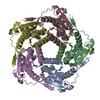
| Entry | Database: PDB / ID: 9g3p | |||||||||||||||||||||||||||||||||
|---|---|---|---|---|---|---|---|---|---|---|---|---|---|---|---|---|---|---|---|---|---|---|---|---|---|---|---|---|---|---|---|---|---|---|
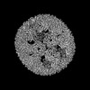
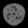
| Title | Circularly permuted lumazine synthase 12-pentamer spherical cage | |||||||||||||||||||||||||||||||||
 Components Components | 6,7-dimethyl-8-ribityllumazine synthase | |||||||||||||||||||||||||||||||||
 Keywords Keywords | DE NOVO PROTEIN / protein cage / protein engineering / self-assembly / geometry / helical reconstruction / bionanotechnology / polymorphism / pentamer | |||||||||||||||||||||||||||||||||
| Function / homology |  Function and homology information Function and homology information6,7-dimethyl-8-ribityllumazine synthase / 6,7-dimethyl-8-ribityllumazine synthase activity / riboflavin synthase complex / riboflavin biosynthetic process / cytosol / cytoplasm Similarity search - Function | |||||||||||||||||||||||||||||||||
| Biological species |   Aquifex aeolicus VF5 (bacteria) Aquifex aeolicus VF5 (bacteria) | |||||||||||||||||||||||||||||||||
| Method | ELECTRON MICROSCOPY / single particle reconstruction / cryo EM / Resolution: 2.08 Å | |||||||||||||||||||||||||||||||||
 Authors Authors | Koziej, L. / Azuma, Y. | |||||||||||||||||||||||||||||||||
| Funding support |  Poland, European Union, 3items Poland, European Union, 3items
| |||||||||||||||||||||||||||||||||
 Citation Citation | Journal: ACS Nano / Year: 2025 Title: Dynamic Assembly of Pentamer-Based Protein Nanotubes. Authors: Lukasz Koziej / Farzad Fatehi / Marta Aleksejczuk / Matthew J Byrne / Jonathan G Heddle / Reidun Twarock / Yusuke Azuma /  Abstract: Hollow proteinaceous particles are useful nanometric containers for delivery and catalysis. Understanding the molecular mechanisms and the geometrical theory behind the polymorphic protein assemblies ...Hollow proteinaceous particles are useful nanometric containers for delivery and catalysis. Understanding the molecular mechanisms and the geometrical theory behind the polymorphic protein assemblies provides a basis for designing ones with the desired morphology. As such, we found that a circularly permuted variant of a cage-forming enzyme, lumazine synthase, cpAaLS, assembles into a variety of hollow spherical and cylindrical structures in response to changes in ionic strength. Cryogenic electron microscopy revealed that these structures are composed entirely of pentameric subunits, and the dramatic cage-to-tube transformation is attributed to the moderately hindered 3-fold symmetry interaction and the imparted torsion angle of the building blocks, where both mechanisms are mediated by an α-helix domain that is untethered from the native position by circular permutation. Mathematical modeling suggests that the unique double- and triple-stranded helical arrangements of subunits are optimal tiling patterns, while different geometries should be possible by modulating the interaction angles of the pentagons. These structural insights into dynamic, pentamer-based protein cages and nanotubes afford guidelines for designing nanoarchitectures with customized morphology and assembly characteristics. | |||||||||||||||||||||||||||||||||
| History |
|
- Structure visualization
| Structure viewer | Molecule: MolmilJmol/JSmol |
|---|
- Downloads & links
-Download
| PDBx/mmCIF format | 9g3p.cif.gz | 1.5 MB | Display | PDBx/mmCIF format |
|---|---|---|---|---|
| PDB format | pdb9g3p.ent.gz | Display | PDB format | |
| PDBx/mmJSON format | 9g3p.json.gz | Tree view | PDBx/mmJSON format | |
| Others |  Other downloads Other downloads |
-Validation report
| Arichive directory | https://data.pdbj.org/pub/pdb/validation_reports/g3/9g3pftp://data.pdbj.org/pub/pdb/validation_reports/g3/9g3p | HTTPS FTP |
|---|
-Related structure data
| Related structure data |  51006MC 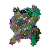 9g3hC 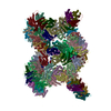 9g3iC 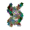 9g3jC 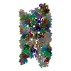 9g3mC 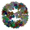 9g3nC 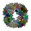 9g3oC M: map data used to model this data C: citing same article ( |
|---|---|
| Similar structure data |
-Links
 PDBj
PDBj
- Assembly
| Deposited unit | 
|
|---|---|
| 1 |
|
-Components
| #1: Protein | Mass: 17449.938 Da / Num. of mol.: 60 Source method: isolated from a genetically manipulated source Details: cpAaLS(84),cpAaLS(84) / Source: (gene. exp.) Aquifex aeolicus VF5 (bacteria) / Gene: ribH, aq_132 / Production host: References: UniProt: O66529, 6,7-dimethyl-8-ribityllumazine synthase Has protein modification | N | |
|---|
-Experimental details
-Experiment
| Experiment | Method: ELECTRON MICROSCOPY |
|---|---|
| EM experiment | Aggregation state: PARTICLE / 3D reconstruction method: single particle reconstruction |
- Sample preparation
| Component | Name: Circularly permuted lumazine synthase 12-pentamer spherical cage Type: COMPLEX Details: Circularly permuted (84) variant of Aquifex aeolicus lumazine synthase Entity ID: all / Source: RECOMBINANT | |||||||||||||||
|---|---|---|---|---|---|---|---|---|---|---|---|---|---|---|---|---|
| Molecular weight |
| |||||||||||||||
| Source (natural) | Organism: Aquifex aeolicus VF5 (bacteria) | |||||||||||||||
| Source (recombinant) | Organism: | |||||||||||||||
| Buffer solution | pH: 8 | |||||||||||||||
| Buffer component |
| |||||||||||||||
| Specimen | Conc.: 1 mg/ml / Embedding applied: NO / Shadowing applied: NO / Staining applied: NO / Vitrification applied: YES | |||||||||||||||
| Specimen support | Grid material: COPPER / Grid mesh size: 200 divisions/in. / Grid type: Quantifoil R2/2 | |||||||||||||||
| Vitrification | Instrument: FEI VITROBOT MARK IV / Cryogen name: ETHANE / Humidity: 100 % / Chamber temperature: 283 K |
- Electron microscopy imaging
| Experimental equipment |  Model: Titan Krios / Image courtesy: FEI Company |
|---|---|
| Microscopy | Model: TFS KRIOS |
| Electron gun | Electron source:  FIELD EMISSION GUN / Accelerating voltage: 300 kV / Illumination mode: FLOOD BEAM FIELD EMISSION GUN / Accelerating voltage: 300 kV / Illumination mode: FLOOD BEAM |
| Electron lens | Mode: BRIGHT FIELD / Calibrated magnification: 105000 X / Nominal defocus max: 2100 nm / Nominal defocus min: 900 nm / Cs: 2.7 mm / C2 aperture diameter: 50 µm / Alignment procedure: COMA FREE |
| Specimen holder | Cryogen: NITROGEN / Specimen holder model: FEI TITAN KRIOS AUTOGRID HOLDER |
| Image recording | Electron dose: 40 e/Å2 / Film or detector model: GATAN K3 BIOCONTINUUM (6k x 4k) / Num. of grids imaged: 1 / Num. of real images: 8486 |
| EM imaging optics | Energyfilter name: GIF Bioquantum / Energyfilter slit width: 20 eV |
| Image scans | Width: 5760 / Height: 4092 |
- Processing
| EM software |
| ||||||||||||||||||||||||||||||||||||||||||||
|---|---|---|---|---|---|---|---|---|---|---|---|---|---|---|---|---|---|---|---|---|---|---|---|---|---|---|---|---|---|---|---|---|---|---|---|---|---|---|---|---|---|---|---|---|---|
| CTF correction | Type: PHASE FLIPPING AND AMPLITUDE CORRECTION | ||||||||||||||||||||||||||||||||||||||||||||
| Particle selection | Num. of particles selected: 590975 / Details: Template picking | ||||||||||||||||||||||||||||||||||||||||||||
| Symmetry | Point symmetry: I (icosahedral) | ||||||||||||||||||||||||||||||||||||||||||||
| 3D reconstruction | Resolution: 2.08 Å / Resolution method: FSC 0.143 CUT-OFF / Num. of particles: 98492 / Algorithm: FOURIER SPACE / Num. of class averages: 1 / Symmetry type: POINT | ||||||||||||||||||||||||||||||||||||||||||||
| Atomic model building | Protocol: RIGID BODY FIT / Space: REAL Details: Initial fitting was done using ChimeraX. Flexible fitting was done using Isolde. | ||||||||||||||||||||||||||||||||||||||||||||
| Atomic model building | PDB-ID: 1HQK Accession code: 1HQK / Source name: PDB / Type: experimental model |