 Movie
Movie Controller

[English] 日本語
 Yorodumi
- EMDB-4328: Structure of a partial yeast 48S preinitiation complex with eIF5 ... -
+ Open data

- Basic information
| Entry | Database: EMDB / ID: EMD-4328 | ||||||||||||
|---|---|---|---|---|---|---|---|---|---|---|---|---|---|
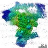
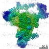
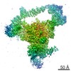
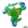
| Title | Structure of a partial yeast 48S preinitiation complex with eIF5 N-terminal domain (Map 1) | ||||||||||||
 Map data Map data | For optimal visualization of all tRNA and eIF2, gauss-filter the map by 1.34 and display it at 0.02 contour level | ||||||||||||
 Sample Sample |
| ||||||||||||
 Keywords Keywords | ribosome / translation / initiation factors / 40S / eIF1A / eIF3 / eIF2 / eIF5 / tRNAi / 48S PIC / small ribosome subunit | ||||||||||||
| Function / homology |  Function and homology information Function and homology informationformation of translation initiation ternary complex / eukaryotic initiation factor eIF2 binding / Cellular response to mitochondrial stress / Recycling of eIF2:GDP / eukaryotic translation initiation factor 3 complex, eIF3e / ABC-family proteins mediated transport / eukaryotic translation initiation factor 3 complex, eIF3m / methionyl-initiator methionine tRNA binding / incipient cellular bud site / translation reinitiation ...formation of translation initiation ternary complex / eukaryotic initiation factor eIF2 binding / Cellular response to mitochondrial stress / Recycling of eIF2:GDP / eukaryotic translation initiation factor 3 complex, eIF3e / ABC-family proteins mediated transport / eukaryotic translation initiation factor 3 complex, eIF3m / methionyl-initiator methionine tRNA binding / incipient cellular bud site / translation reinitiation / eukaryotic translation initiation factor 2 complex / eukaryotic translation initiation factor 3 complex / formation of cytoplasmic translation initiation complex / cytoplasmic translational initiation / multi-eIF complex / protein-synthesizing GTPase / eukaryotic 43S preinitiation complex / formation of translation preinitiation complex / eukaryotic 48S preinitiation complex / positive regulation of translational fidelity / GDP-dissociation inhibitor activity / regulation of translational initiation / Formation of the ternary complex, and subsequently, the 43S complex / Translation initiation complex formation / Ribosomal scanning and start codon recognition / Formation of a pool of free 40S subunits / L13a-mediated translational silencing of Ceruloplasmin expression / ribosomal small subunit binding / endonucleolytic cleavage to generate mature 3'-end of SSU-rRNA from (SSU-rRNA, 5.8S rRNA, LSU-rRNA) / 90S preribosome / translation regulator activity / endonucleolytic cleavage in ITS1 to separate SSU-rRNA from 5.8S rRNA and LSU-rRNA from tricistronic rRNA transcript (SSU-rRNA, 5.8S rRNA, LSU-rRNA) / negative regulation of translational initiation / translation initiation factor binding / translation initiation factor activity / GTPase activator activity / rescue of stalled ribosome / maturation of SSU-rRNA from tricistronic rRNA transcript (SSU-rRNA, 5.8S rRNA, LSU-rRNA) / cytosolic ribosome assembly / maturation of SSU-rRNA / small-subunit processome / translational initiation / protein kinase C binding / positive regulation of apoptotic signaling pathway / cytoplasmic stress granule / modification-dependent protein catabolic process / rRNA processing / protein tag activity / double-stranded RNA binding / ribosomal small subunit biogenesis / small ribosomal subunit rRNA binding / ribosome binding / ribosomal small subunit assembly / small ribosomal subunit / cytosolic small ribosomal subunit / cytosolic large ribosomal subunit / cytoplasmic translation / rRNA binding / ribosome / protein ubiquitination / structural constituent of ribosome / ribonucleoprotein complex / translation / positive regulation of protein phosphorylation / GTPase activity / mRNA binding / ubiquitin protein ligase binding / GTP binding / nucleolus / protein kinase binding / RNA binding / zinc ion binding / identical protein binding / nucleus / metal ion binding / cytosol / cytoplasm Similarity search - Function | ||||||||||||
| Biological species |  Kluyveromyces lactis (strain ATCC 8585 / CBS 2359 / DSM 70799 / NBRC 1267 / NRRL Y-1140 / WM37) (yeast) / Kluyveromyces lactis (strain ATCC 8585 / CBS 2359 / DSM 70799 / NBRC 1267 / NRRL Y-1140 / WM37) (yeast) / | ||||||||||||
| Method | single particle reconstruction / cryo EM / Resolution: 3.02 Å | ||||||||||||
 Authors Authors | Llacer JL / Hussain T | ||||||||||||
| Funding support |  United Kingdom, 3 items United Kingdom, 3 items
| ||||||||||||
 Citation Citation | Journal: Elife / Year: 2018 Title: Translational initiation factor eIF5 replaces eIF1 on the 40S ribosomal subunit to promote start-codon recognition. Authors: Jose Luis Llácer / Tanweer Hussain / Adesh K Saini / Jagpreet Singh Nanda / Sukhvir Kaur / Yuliya Gordiyenko / Rakesh Kumar / Alan G Hinnebusch / Jon R Lorsch / V Ramakrishnan /    Abstract: In eukaryotic translation initiation, AUG recognition of the mRNA requires accommodation of Met-tRNA in a 'P' state, which is antagonized by the factor eIF1. eIF5 is a GTPase activating protein (GAP) ...In eukaryotic translation initiation, AUG recognition of the mRNA requires accommodation of Met-tRNA in a 'P' state, which is antagonized by the factor eIF1. eIF5 is a GTPase activating protein (GAP) of eIF2 that additionally promotes stringent AUG selection, but the molecular basis of its dual function was unknown. We present a cryo-electron microscopy (cryo-EM) reconstruction of a yeast 48S pre-initiation complex (PIC), at an overall resolution of 3.0 Å, featuring the N-terminal domain (NTD) of eIF5 bound to the 40S subunit at the location vacated by eIF1. eIF5 interacts with and allows a more accommodated orientation of Met-tRNA. Substitutions of eIF5 residues involved in the eIF5-NTD/tRNA interaction influenced initiation at near-cognate UUG codons and the closed/open PIC conformation in vitro, consistent with direct stabilization of the codon:anticodon duplex by the wild-type eIF5-NTD. The present structure reveals the basis for a key role of eIF5 in start-codon selection. | ||||||||||||
| History |
|
- Structure visualization
| Movie |
Movie viewer |
|---|---|
| Structure viewer | EM map: SurfViewMolmilJmol/JSmol |
| Supplemental images |

- Downloads & links
-EMDB archive
| Map data | emd_4328.map.gz | 95.8 MB | EMDB map data format | |
|---|---|---|---|---|
| Header (meta data) | emd-4328-v30.xmlemd-4328.xml | 85.5 KB 85.5 KB | Display Display | EMDB header |
| Images | 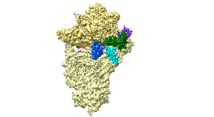 emd_4328.png emd_4328.png | 188.5 KB | ||
| Filedesc metadata | emd-4328.cif.gz | 16.9 KB | ||
| Others | emd_4328_half_map_1.map.gzemd_4328_half_map_2.map.gz | 80.7 MB 80.7 MB | ||
| Archive directory |  http://ftp.pdbj.org/pub/emdb/structures/EMD-4328ftp://ftp.pdbj.org/pub/emdb/structures/EMD-4328 http://ftp.pdbj.org/pub/emdb/structures/EMD-4328ftp://ftp.pdbj.org/pub/emdb/structures/EMD-4328 | HTTPS FTP |
-Validation report
| Summary document | emd_4328_validation.pdf.gz | 919.6 KB | Display | EMDB validaton report |
|---|---|---|---|---|
| Full document | emd_4328_full_validation.pdf.gz | 919.2 KB | Display | |
| Data in XML | emd_4328_validation.xml.gz | 13.3 KB | Display | |
| Data in CIF | emd_4328_validation.cif.gz | 15.5 KB | Display | |
| Arichive directory | https://ftp.pdbj.org/pub/emdb/validation_reports/EMD-4328ftp://ftp.pdbj.org/pub/emdb/validation_reports/EMD-4328 | HTTPS FTP |
-Related structure data
| Related structure data |  6fyyMC  4327C  4329C 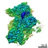 4330C 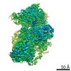 4331C 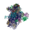 6fyxC C: citing same article ( M: atomic model generated by this map |
|---|---|
| Similar structure data |


-Links
| EMDB pages | EMDB (EBI/PDBe) / EMDataResource |
|---|---|
| Related items in Molecule of the Month |


-Map
| File | Download / File: emd_4328.map.gz / Format: CCP4 / Size: 103 MB / Type: IMAGE STORED AS FLOATING POINT NUMBER (4 BYTES) | ||||||||||||||||||||||||||||||||||||||||||||||||||||||||||||||||||||
|---|---|---|---|---|---|---|---|---|---|---|---|---|---|---|---|---|---|---|---|---|---|---|---|---|---|---|---|---|---|---|---|---|---|---|---|---|---|---|---|---|---|---|---|---|---|---|---|---|---|---|---|---|---|---|---|---|---|---|---|---|---|---|---|---|---|---|---|---|---|
| Annotation | For optimal visualization of all tRNA and eIF2, gauss-filter the map by 1.34 and display it at 0.02 contour level | ||||||||||||||||||||||||||||||||||||||||||||||||||||||||||||||||||||
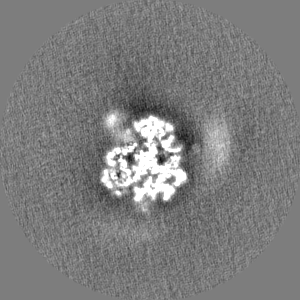
| Projections & slices | Image control
Images are generated by Spider. | ||||||||||||||||||||||||||||||||||||||||||||||||||||||||||||||||||||
| Voxel size | X=Y=Z: 1.34 Å | ||||||||||||||||||||||||||||||||||||||||||||||||||||||||||||||||||||
| Density |
| ||||||||||||||||||||||||||||||||||||||||||||||||||||||||||||||||||||
| Symmetry | Space group: 1 | ||||||||||||||||||||||||||||||||||||||||||||||||||||||||||||||||||||
| Details | EMDB XML:
CCP4 map header:
| ||||||||||||||||||||||||||||||||||||||||||||||||||||||||||||||||||||
 Z (Sec.)
 Y (Row.)
 X (Col.)


-Supplemental data
-Half map: #1
| File | emd_4328_half_map_1.map | ||||||||||||
|---|---|---|---|---|---|---|---|---|---|---|---|---|---|
| Projections & Slices |
| ||||||||||||
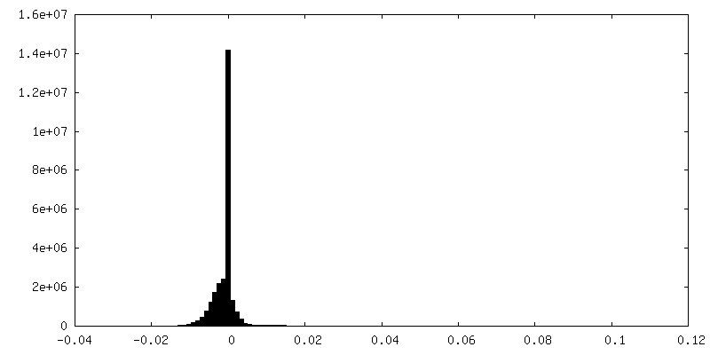
| Density Histograms |


-Half map: #2
| File | emd_4328_half_map_2.map | ||||||||||||
|---|---|---|---|---|---|---|---|---|---|---|---|---|---|
| Projections & Slices |
| ||||||||||||
| Density Histograms |


- Sample components
+Entire : Structure of a partial yeast 48S preinitiation complex with eIF5 ...
+Supramolecule #1: Structure of a partial yeast 48S preinitiation complex with eIF5 ...
+Supramolecule #2: Ribosome
+Supramolecule #3: tRNA
+Supramolecule #4: initiation factors
+Supramolecule #5: initiation factors
+Supramolecule #6: mRNA
+Macromolecule #1: tRNAi
+Macromolecule #2: 18S ribosomal RNA
+Macromolecule #3: mRNA (31-MER)
+Macromolecule #4: 40S ribosomal protein S0
+Macromolecule #5: 40S ribosomal protein S1
+Macromolecule #6: KLLA0F09812p
+Macromolecule #7: KLLA0D08305p
+Macromolecule #8: 40S ribosomal protein S4
+Macromolecule #9: KLLA0D10659p
+Macromolecule #10: 40S ribosomal protein S6
+Macromolecule #11: 40S ribosomal protein S7
+Macromolecule #12: 40S ribosomal protein S8
+Macromolecule #13: KLLA0E23673p
+Macromolecule #14: KLLA0B08173p
+Macromolecule #15: KLLA0A10483p
+Macromolecule #16: 40S ribosomal protein S12
+Macromolecule #17: KLLA0F18040p
+Macromolecule #18: 40S ribosomal protein S14
+Macromolecule #19: KLLA0F07843p
+Macromolecule #20: 40S ribosomal protein S16
+Macromolecule #21: KLLA0B01474p
+Macromolecule #22: KLLA0B01562p
+Macromolecule #23: KLLA0A07194p
+Macromolecule #24: KLLA0F25542p
+Macromolecule #25: 40S ribosomal protein S21
+Macromolecule #26: 40S ribosomal protein S22
+Macromolecule #27: KLLA0B11231p
+Macromolecule #28: 40S ribosomal protein S24
+Macromolecule #29: KLLA0B06182p
+Macromolecule #30: 40S ribosomal protein S26
+Macromolecule #31: 40S ribosomal protein S27
+Macromolecule #32: 40S ribosomal protein S28
+Macromolecule #33: 40S ribosomal protein S29
+Macromolecule #34: 40S ribosomal protein S30
+Macromolecule #35: Ubiquitin-40S ribosomal protein S27a
+Macromolecule #36: KLLA0E12277p
+Macromolecule #37: 60S ribosomal protein L41-A
+Macromolecule #38: Eukaryotic translation initiation factor 1A

+Macromolecule #39: Eukaryotic translation initiation factor 2 subunit alpha
+Macromolecule #40: Eukaryotic translation initiation factor 2 subunit gamma
+Macromolecule #41: Eukaryotic translation initiation factor 2 subunit beta
+Macromolecule #42: Eukaryotic translation initiation factor 5
+Macromolecule #43: Eukaryotic translation initiation factor 3 subunit A
+Macromolecule #44: Eukaryotic translation initiation factor 3 subunit B
+Macromolecule #45: eIF3c,Eukaryotic translation initiation factor 3 subunit C
+Macromolecule #46: Eukaryotic translation initiation factor 3 subunit G
+Macromolecule #47: Eukaryotic translation initiation factor 3 subunit I
+Macromolecule #48: MAGNESIUM ION
+Macromolecule #49: ZINC ION
+Macromolecule #50: METHIONINE

+Macromolecule #51: PHOSPHOMETHYLPHOSPHONIC ACID GUANYLATE ESTER

-Experimental details
-Structure determination
| Method | cryo EM |
|---|---|
 Processing Processing | single particle reconstruction |
| Aggregation state | particle |
-Sample preparation
| Concentration | 0.15 mg/mL | ||||||||||||||||||
|---|---|---|---|---|---|---|---|---|---|---|---|---|---|---|---|---|---|---|---|
| Buffer | pH: 6.5 Component:
| ||||||||||||||||||
| Grid | Model: Quantifoil R2/2 / Material: COPPER / Mesh: 400 / Support film - Material: CARBON / Support film - topology: CONTINUOUS / Pretreatment - Type: GLOW DISCHARGE / Pretreatment - Time: 20 sec. | ||||||||||||||||||
| Vitrification | Cryogen name: ETHANE / Chamber humidity: 100 % / Chamber temperature: 277 K / Instrument: FEI VITROBOT MARK I |
- Electron microscopy
| Microscope | FEI POLARA 300 |
|---|---|
| Temperature | Min: 90.0 K / Max: 100.0 K |
| Image recording | Film or detector model: FEI FALCON III (4k x 4k) / Detector mode: INTEGRATING / Number grids imaged: 2 / Number real images: 2100 / Average exposure time: 1.1 sec. / Average electron dose: 40.0 e/Å2 Details: Images were collected in movie-mode at 32 frames per second |
| Electron beam | Acceleration voltage: 300 kV / Electron source:  FIELD EMISSION GUN FIELD EMISSION GUN |
| Electron optics | C2 aperture diameter: 50.0 µm / Calibrated magnification: 104478 / Illumination mode: FLOOD BEAM / Imaging mode: BRIGHT FIELD / Cs: 2.0 mm / Nominal defocus max: 3.5 µm / Nominal defocus min: 1.5 µm / Nominal magnification: 78000 |
| Sample stage | Cooling holder cryogen: NITROGEN |
| Experimental equipment |  Model: Tecnai Polara / Image courtesy: FEI Company |
+Image processing
-Atomic model buiding 1
| Refinement | Space: RECIPROCAL / Protocol: OTHER / Overall B value: 49 / Target criteria: FSC |
|---|---|
| Output model | PDB-6fyy: |
