Movie
Movie Controller

[English] 日本語
 Yorodumi
- EMDB-0685: Cryo-EM structure of the catalytic activated yeast spliceosome (B... -
+ Open data

- Basic information
| Entry | Database: EMDB / ID: EMD-0685 | |||||||||
|---|---|---|---|---|---|---|---|---|---|---|
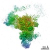
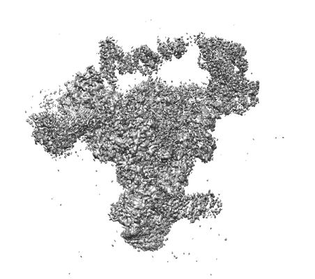
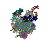
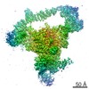
| Title | Cryo-EM structure of the catalytic activated yeast spliceosome (B* complex) assembled on UBC4 pre-mRNA at 3.2 angstrom resolution | |||||||||
 Map data Map data | ||||||||||
 Sample Sample |
| |||||||||
| Biological species |  | |||||||||
| Method | single particle reconstruction / cryo EM / Resolution: 3.2 Å | |||||||||
 Authors Authors | Wan R / Bai R / Yan C / Lei J / Shi Y | |||||||||
 Citation Citation | Journal: Cell / Year: 2019 Title: Structures of the Catalytically Activated Yeast Spliceosome Reveal the Mechanism of Branching. Authors: Ruixue Wan / Rui Bai / Chuangye Yan / Jianlin Lei / Yigong Shi /  Abstract: Pre-mRNA splicing is executed by the spliceosome. Structural characterization of the catalytically activated complex (B) is pivotal for understanding the branching reaction. In this study, we ...Pre-mRNA splicing is executed by the spliceosome. Structural characterization of the catalytically activated complex (B) is pivotal for understanding the branching reaction. In this study, we assembled the B complexes on two different pre-mRNAs from Saccharomyces cerevisiae and determined the cryo-EM structures of four distinct B complexes at overall resolutions of 2.9-3.8 Å. The duplex between U2 small nuclear RNA (snRNA) and the branch point sequence (BPS) is discretely away from the 5'-splice site (5'SS) in the three B complexes that are devoid of the step I splicing factors Yju2 and Cwc25. Recruitment of Yju2 into the active site brings the U2/BPS duplex into the vicinity of 5'SS, with the BPS nucleophile positioned 4 Å away from the catalytic metal M2. This analysis reveals the functional mechanism of Yju2 and Cwc25 in branching. These structures on different pre-mRNAs reveal substrate-specific conformations of the spliceosome in a major functional state. | |||||||||
| History |
|
- Structure visualization
| Movie |
 Movie viewer Movie viewer |
|---|---|
| Structure viewer | EM map: SurfViewMolmilJmol/JSmol |
| Supplemental images |
- Downloads & links
-EMDB archive
| Map data | emd_0685.map.gz | 224.6 MB | EMDB map data format | |
|---|---|---|---|---|
| Header (meta data) | emd-0685-v30.xmlemd-0685.xml | 8.4 KB 8.4 KB | Display Display | EMDB header |
| Images |  emd_0685.png emd_0685.png | 57.3 KB | ||
| Archive directory |  http://ftp.pdbj.org/pub/emdb/structures/EMD-0685ftp://ftp.pdbj.org/pub/emdb/structures/EMD-0685 http://ftp.pdbj.org/pub/emdb/structures/EMD-0685ftp://ftp.pdbj.org/pub/emdb/structures/EMD-0685 | HTTPS FTP |
-Related structure data
| Related structure data | 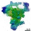 0684C 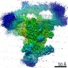 0686C 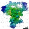 0687C 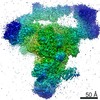 0691C 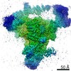 0692C 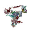 6j6gC 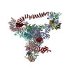 6j6hC  6j6nC 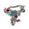 6j6qC C: citing same article ( |
|---|---|
| Similar structure data |


-Links
| EMDB pages | EMDB (EBI/PDBe) / EMDataResource |
|---|
-Map
| File | Download / File: emd_0685.map.gz / Format: CCP4 / Size: 244.1 MB / Type: IMAGE STORED AS FLOATING POINT NUMBER (4 BYTES) | ||||||||||||||||||||||||||||||||||||||||||||||||||||||||||||
|---|---|---|---|---|---|---|---|---|---|---|---|---|---|---|---|---|---|---|---|---|---|---|---|---|---|---|---|---|---|---|---|---|---|---|---|---|---|---|---|---|---|---|---|---|---|---|---|---|---|---|---|---|---|---|---|---|---|---|---|---|---|
| Projections & slices | Image control
Images are generated by Spider. | ||||||||||||||||||||||||||||||||||||||||||||||||||||||||||||
| Voxel size | X=Y=Z: 1.33 Å | ||||||||||||||||||||||||||||||||||||||||||||||||||||||||||||
| Density |
| ||||||||||||||||||||||||||||||||||||||||||||||||||||||||||||
| Symmetry | Space group: 1 | ||||||||||||||||||||||||||||||||||||||||||||||||||||||||||||
| Details | EMDB XML:
CCP4 map header:
| ||||||||||||||||||||||||||||||||||||||||||||||||||||||||||||
 Z (Sec.)
 Y (Row.)
 X (Col.)


-Supplemental data
- Sample components
-Entire : the catalytic activated spliceosome B* complex assembled on UBC4 ...
| Entire | Name: the catalytic activated spliceosome B* complex assembled on UBC4 pre-mRNA |
|---|---|
| Components |
|
-Supramolecule #1: the catalytic activated spliceosome B* complex assembled on UBC4 ...
| Supramolecule | Name: the catalytic activated spliceosome B* complex assembled on UBC4 pre-mRNA type: complex / ID: 1 / Parent: 0 |
|---|---|
| Source (natural) | Organism: |
-Experimental details
-Structure determination
| Method | cryo EM |
|---|---|
 Processing Processing | single particle reconstruction |
| Aggregation state | particle |
-Sample preparation
| Buffer | pH: 7.9 |
|---|---|
| Vitrification | Cryogen name: ETHANE |
- Electron microscopy
| Microscope | FEI TITAN KRIOS |
|---|---|
| Image recording | Film or detector model: GATAN K2 SUMMIT (4k x 4k) / Detector mode: SUPER-RESOLUTION / Average electron dose: 49.3 e/Å2 |
| Electron beam | Acceleration voltage: 300 kV / Electron source:  FIELD EMISSION GUN FIELD EMISSION GUN |
| Electron optics | Illumination mode: FLOOD BEAM / Imaging mode: BRIGHT FIELD |
| Experimental equipment |  Model: Titan Krios / Image courtesy: FEI Company |