 Movie
Movie Controller


+ Open data

- Basic information
| Entry | Database: EMDB / ID: EMD-4240 | |||||||||
|---|---|---|---|---|---|---|---|---|---|---|
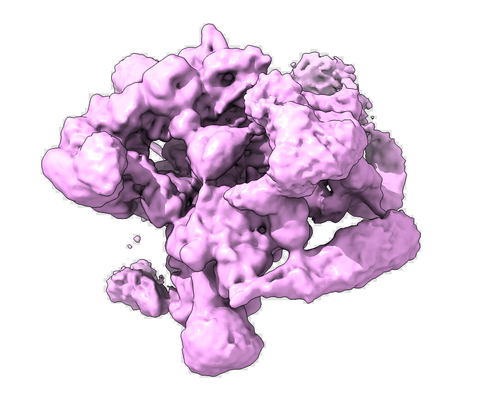
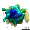
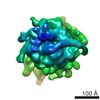
| Title | Human Bact spliceosome state 8 unmasked | |||||||||
 Map data Map data | ||||||||||
 Sample Sample |
| |||||||||
| Function / homology |  Function and homology information Function and homology informationRES complex / post-spliceosomal complex / negative regulation of chemokine-mediated signaling pathway / regulation of vitamin D receptor signaling pathway / oocyte development / U11/U12 snRNP / regulation of retinoic acid receptor signaling pathway / blastocyst development / post-mRNA release spliceosomal complex / U2 snRNP binding ...RES complex / post-spliceosomal complex / negative regulation of chemokine-mediated signaling pathway / regulation of vitamin D receptor signaling pathway / oocyte development / U11/U12 snRNP / regulation of retinoic acid receptor signaling pathway / blastocyst development / post-mRNA release spliceosomal complex / U2 snRNP binding / U7 snRNA binding / histone pre-mRNA DCP binding / generation of catalytic spliceosome for first transesterification step / U7 snRNP / blastocyst formation / 3'-5' RNA helicase activity / cis assembly of pre-catalytic spliceosome / histone pre-mRNA 3'end processing complex / nuclear histone mRNA catabolic process / SLBP independent Processing of Histone Pre-mRNAs / SLBP Dependent Processing of Replication-Dependent Histone Pre-mRNAs / inner cell mass cell proliferation / miRNA processing / alternative mRNA splicing, via spliceosome / poly(A) binding / 7-methylguanosine cap hypermethylation / U12-type spliceosomal complex / nuclear retinoic acid receptor binding / embryonic brain development / positive regulation of mRNA splicing, via spliceosome / U1 snRNP binding / C2H2 zinc finger domain binding / pICln-Sm protein complex / regulation of mRNA splicing, via spliceosome / U2-type catalytic step 1 spliceosome / methylosome / RNA splicing, via transesterification reactions / mRNA 3'-end processing / sno(s)RNA-containing ribonucleoprotein complex / small nuclear ribonucleoprotein complex / SMN-Sm protein complex / protein methylation / splicing factor binding / spliceosomal tri-snRNP complex / positive regulation of vitamin D receptor signaling pathway / P granule / commitment complex / U4 snRNP / snRNP binding / host-mediated activation of viral transcription / U2-type precatalytic spliceosome / mRNA cis splicing, via spliceosome / Regulation of gene expression in late stage (branching morphogenesis) pancreatic bud precursor cells / RUNX3 regulates NOTCH signaling / Notch binding / U2-type prespliceosome assembly / U2-type catalytic step 2 spliceosome / U2-type spliceosomal complex / nuclear vitamin D receptor binding / Transport of Mature mRNA derived from an Intron-Containing Transcript / NOTCH4 Intracellular Domain Regulates Transcription / telomerase holoenzyme complex / telomerase RNA binding / pre-mRNA binding / positive regulation of neurogenesis / spliceosome conformational change to release U4 (or U4atac) and U1 (or U11) / U1 snRNP / U2 snRNP / RNA Polymerase II Transcription Termination / NOTCH3 Intracellular Domain Regulates Transcription / mRNA stabilization / U2-type prespliceosome / RHOBTB1 GTPase cycle / K63-linked polyubiquitin modification-dependent protein binding / protein peptidyl-prolyl isomerization / nuclear androgen receptor binding / cyclosporin A binding / generation of catalytic spliceosome for second transesterification step / precatalytic spliceosome / ubiquitin-ubiquitin ligase activity / Notch-HLH transcription pathway / Formation of paraxial mesoderm / lipid biosynthetic process / pattern recognition receptor activity / WD40-repeat domain binding / positive regulation of transforming growth factor beta receptor signaling pathway / mRNA 3'-splice site recognition / regulation of RNA splicing / SMAD binding / positive regulation of transcription by RNA polymerase I / mRNA Splicing - Minor Pathway / positive regulation of transcription by RNA polymerase III / spliceosomal complex assembly / intrinsic apoptotic signaling pathway in response to DNA damage by p53 class mediator / Prp19 complex / negative regulation of mRNA splicing, via spliceosome / spliceosomal tri-snRNP complex assembly / U5 snRNP / U5 snRNA binding / ovarian follicle development Similarity search - Function | |||||||||
| Biological species |  Homo sapiens (human) Homo sapiens (human) | |||||||||
| Method | single particle reconstruction / cryo EM / Resolution: 4.5 Å | |||||||||
 Authors Authors | Haselbach D / Komarov I / Agafonov D / Kastner B / Luehrmann R / Stark H | |||||||||
 Citation Citation | Journal: Cell / Year: 2018 Title: Structure and Conformational Dynamics of the Human Spliceosomal B Complex. Authors: David Haselbach / Ilya Komarov / Dmitry E Agafonov / Klaus Hartmuth / Benjamin Graf / Olexandr Dybkov / Henning Urlaub / Berthold Kastner / Reinhard Lührmann / Holger Stark /  Abstract: The spliceosome is a highly dynamic macromolecular complex that precisely excises introns from pre-mRNA. Here we report the cryo-EM 3D structure of the human B spliceosome at 3.4 Å resolution. In ...The spliceosome is a highly dynamic macromolecular complex that precisely excises introns from pre-mRNA. Here we report the cryo-EM 3D structure of the human B spliceosome at 3.4 Å resolution. In the B state, the spliceosome is activated but not catalytically primed, so that it is functionally blocked prior to the first catalytic step of splicing. The spliceosomal core is similar to the yeast B spliceosome; important differences include the presence of the RNA helicase aquarius and peptidyl prolyl isomerases. To examine the overall dynamic behavior of the purified spliceosome, we developed a principal component analysis-based approach. Calculating the energy landscape revealed eight major conformational states, which we refined to higher resolution. Conformational differences of the highly flexible structural components between these eight states reveal how spliceosomal components contribute to the assembly of the spliceosome, allowing it to generate a dynamic interaction network required for its subsequent catalytic activation. | |||||||||
| History |
|
- Structure visualization
| Movie |
Movie viewer |
|---|---|
| Structure viewer | EM map: SurfViewMolmilJmol/JSmol |
| Supplemental images |
- Downloads & links
-EMDB archive
| Map data | emd_4240.map.gz | 245.3 MB | EMDB map data format | |
|---|---|---|---|---|
| Header (meta data) | emd-4240-v30.xmlemd-4240.xml | 13.8 KB 13.8 KB | Display Display | EMDB header |
| Images |  emd_4240.png emd_4240.png | 144.4 KB | ||
| Archive directory |  http://ftp.pdbj.org/pub/emdb/structures/EMD-4240ftp://ftp.pdbj.org/pub/emdb/structures/EMD-4240 http://ftp.pdbj.org/pub/emdb/structures/EMD-4240ftp://ftp.pdbj.org/pub/emdb/structures/EMD-4240 | HTTPS FTP |
-Related structure data
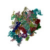
| Related structure data |  6ff7MC  4233C  4234C 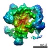 4235C 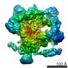 4236C 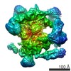 4237C 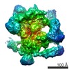 4238C 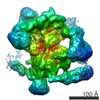 4239C 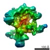 4247C 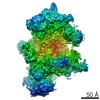 4248C 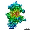 4249C 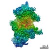 4250C 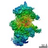 4251C 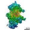 4252C 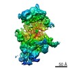 4253C 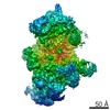 4254C 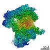 4255C 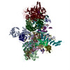 6ff4C C: citing same article ( M: atomic model generated by this map |
|---|---|
| Similar structure data | |
| EM raw data | EMPIAR-10160 (Title: Conformational Dynamics of human Bact spliceosome / Data size: 2.8 TB Data #1: aligned and summed micrograph stack of human Bact spliceosome [micrographs - single frame] Data #2: aligned, dose-weighted and summed micrograph stack of human Bact spliceosome [micrographs - single frame]) |

-Links
| EMDB pages | EMDB (EBI/PDBe) / EMDataResource |
|---|---|
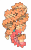
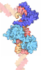
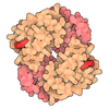
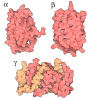
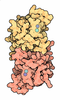
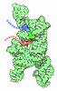
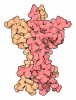
| Related items in Molecule of the Month |













-Map
| File | Download / File: emd_4240.map.gz / Format: CCP4 / Size: 282.6 MB / Type: IMAGE STORED AS FLOATING POINT NUMBER (4 BYTES) | ||||||||||||||||||||||||||||||||||||||||||||||||||||||||||||||||||||
|---|---|---|---|---|---|---|---|---|---|---|---|---|---|---|---|---|---|---|---|---|---|---|---|---|---|---|---|---|---|---|---|---|---|---|---|---|---|---|---|---|---|---|---|---|---|---|---|---|---|---|---|---|---|---|---|---|---|---|---|---|---|---|---|---|---|---|---|---|---|
| Projections & slices | Image control
Images are generated by Spider. | ||||||||||||||||||||||||||||||||||||||||||||||||||||||||||||||||||||
| Voxel size | X=Y=Z: 1.16 Å | ||||||||||||||||||||||||||||||||||||||||||||||||||||||||||||||||||||
| Density |
| ||||||||||||||||||||||||||||||||||||||||||||||||||||||||||||||||||||
| Symmetry | Space group: 1 | ||||||||||||||||||||||||||||||||||||||||||||||||||||||||||||||||||||
| Details | EMDB XML:
CCP4 map header:
| ||||||||||||||||||||||||||||||||||||||||||||||||||||||||||||||||||||
 Z (Sec.)
 Y (Row.)
 X (Col.)


-Supplemental data
- Sample components
-Entire : human Bact spliceosome state 1 unmasked
| Entire | Name: human Bact spliceosome state 1 unmasked |
|---|---|
| Components |
|
-Supramolecule #1: human Bact spliceosome state 1 unmasked
| Supramolecule | Name: human Bact spliceosome state 1 unmasked / type: complex / ID: 1 / Parent: 0 |
|---|---|
| Source (natural) | Organism: Homo sapiens (human) / Strain: HELA |
| Molecular weight | Theoretical: 4.5 MDa |
-Experimental details
-Structure determination
| Method | cryo EM |
|---|---|
 Processing Processing | single particle reconstruction |
| Aggregation state | particle |
-Sample preparation
| Concentration | 0.05 mg/mL | ||||||||||||
|---|---|---|---|---|---|---|---|---|---|---|---|---|---|
| Buffer | pH: 7.9 Component:
| ||||||||||||
| Grid | Model: Quantifoil R3.5/1 / Material: COPPER / Mesh: 400 / Support film - #0 - Film type ID: 1 / Support film - #0 - Material: CARBON / Support film - #0 - topology: HOLEY / Support film - #1 - Film type ID: 2 / Support film - #1 - Material: CARBON / Support film - #1 - topology: CONTINUOUS | ||||||||||||
| Vitrification | Cryogen name: ETHANE / Chamber humidity: 75 % / Chamber temperature: 277 K / Instrument: LEICA EM GP / Details: blot with blotting sensor. |
- Electron microscopy
| Microscope | FEI TITAN KRIOS |
|---|---|
| Alignment procedure | Coma free - Residual tilt: 14.0 mrad |
| Specialist optics | Spherical aberration corrector: Microscope was modified with a Cs corrector with two hexapoles elements |
| Image recording | Film or detector model: FEI FALCON III (4k x 4k) / Detector mode: INTEGRATING / Digitization - Dimensions - Width: 4096 pixel / Digitization - Dimensions - Height: 4096 pixel / Digitization - Sampling interval: 14.0 µm / Number grids imaged: 1 / Number real images: 32000 / Average exposure time: 1.0 sec. / Average electron dose: 40.0 e/Å2 |
| Electron beam | Acceleration voltage: 300 kV / Electron source:  FIELD EMISSION GUN FIELD EMISSION GUN |
| Electron optics | C2 aperture diameter: 70.0 µm / Illumination mode: SPOT SCAN / Imaging mode: BRIGHT FIELD / Cs: 0.001 mm / Nominal defocus max: 4.5 µm / Nominal defocus min: 0.8 µm / Nominal magnification: 59000 |
| Sample stage | Specimen holder model: FEI TITAN KRIOS AUTOGRID HOLDER / Cooling holder cryogen: NITROGEN |
| Experimental equipment |  Model: Titan Krios / Image courtesy: FEI Company |
+Image processing
-Atomic model buiding 1
| Refinement | Protocol: RIGID BODY FIT |
|---|---|
| Output model | PDB-6ff7: |
