Movie
Movie Controller

[English] 日本語
 Yorodumi
- EMDB-41411: Structural architecture of the acidic region of the B domain of c... -
+ Open data

- Basic information
| Entry |  | ||||||||||||||||||
|---|---|---|---|---|---|---|---|---|---|---|---|---|---|---|---|---|---|---|---|
| Title | Structural architecture of the acidic region of the B domain of coagulation factor V | ||||||||||||||||||
 Map data Map data | Coagulation plasma factor V - Sharpened composite map | ||||||||||||||||||
 Sample Sample |
| ||||||||||||||||||
 Keywords Keywords | Coagulation / Pro-cofactor / Factor V / B Domain / Acidic Region / BLOOD CLOTTING | ||||||||||||||||||
| Function / homology |  Function and homology information Function and homology informationresponse to vitamin K / platelet alpha granule / Cargo concentration in the ER / COPII-coated ER to Golgi transport vesicle / COPII-mediated vesicle transport / blood circulation / : / endoplasmic reticulum-Golgi intermediate compartment membrane / platelet alpha granule lumen / Post-translational protein phosphorylation ...response to vitamin K / platelet alpha granule / Cargo concentration in the ER / COPII-coated ER to Golgi transport vesicle / COPII-mediated vesicle transport / blood circulation / : / endoplasmic reticulum-Golgi intermediate compartment membrane / platelet alpha granule lumen / Post-translational protein phosphorylation / Regulation of Insulin-like Growth Factor (IGF) transport and uptake by Insulin-like Growth Factor Binding Proteins (IGFBPs) / blood coagulation / Platelet degranulation / extracellular vesicle / endoplasmic reticulum lumen / copper ion binding / : / extracellular region / membrane / plasma membrane Similarity search - Function | ||||||||||||||||||
| Biological species |  Homo sapiens (human) Homo sapiens (human) | ||||||||||||||||||
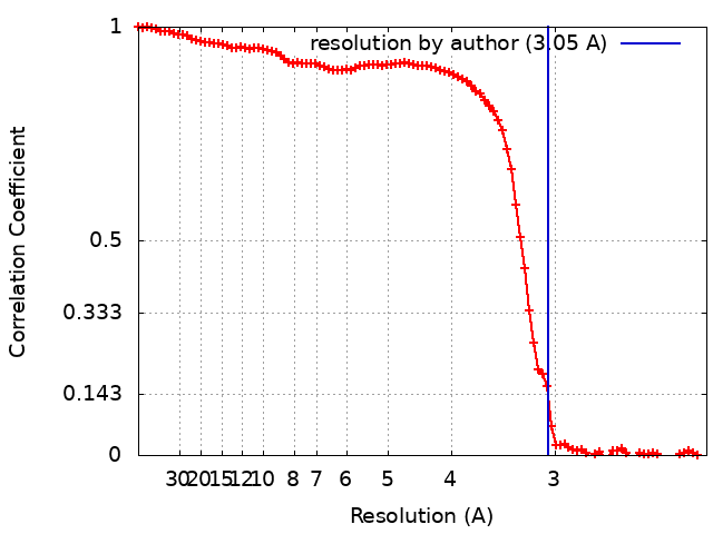
| Method | single particle reconstruction / cryo EM / Resolution: 3.05 Å | ||||||||||||||||||
 Authors Authors | Mohammed BM / Basore K / Summers B / Pelc LA / Di Cera E | ||||||||||||||||||
| Funding support |  United States, 5 items United States, 5 items
| ||||||||||||||||||
 Citation Citation | Journal: J Thromb Haemost / Year: 2024 Title: Structural architecture of the acidic region of the B domain of coagulation factor V. Authors: Bassem M Mohammed / Katherine Basore / Brock Summers / Leslie A Pelc / Enrico Di Cera / Abstract: BACKGROUND: Coagulation factor (F)V features an A1-A2-B-A3-C1-C2 domain organization and functions as the inactive precursor of FVa, a component of the prothrombinase complex required for rapid ...BACKGROUND: Coagulation factor (F)V features an A1-A2-B-A3-C1-C2 domain organization and functions as the inactive precursor of FVa, a component of the prothrombinase complex required for rapid thrombin generation in the penultimate step of the coagulation cascade. An intramolecular interaction within the large B domain (residues 710-1545) involves the basic region (BR, residues 963-1008) and acidic region (AR, residues 1493-1537) and locks FV in its inactive state. However, structural information on this important regulatory interaction or on the separate architecture of the AR and BR remains elusive due to conformational disorder of the B domain. OBJECTIVES: To reveal the structure of the BR-AR interaction or of its separate components. METHODS: The structure of FV is solved by cryogenic electron microscopy. RESULTS: A new 3.05 Å resolution cryogenic electron microscopy structure of FV confirms the overall organization of the A and C domains but resolves the segment 1507 to 1545 within a largely ...RESULTS: A new 3.05 Å resolution cryogenic electron microscopy structure of FV confirms the overall organization of the A and C domains but resolves the segment 1507 to 1545 within a largely disordered B domain. The segment contains most of the AR and is organized as recently reported in FV short, a spliced variant of FV with a significantly shorter and less disordered B domain. CONCLUSION: The similar architecture of the AR in FV and FV short provides structural context for physiologically important interactions of this region with the BR in FV and with the basic C-terminal ...CONCLUSION: The similar architecture of the AR in FV and FV short provides structural context for physiologically important interactions of this region with the BR in FV and with the basic C-terminal end of tissue factor pathway inhibitor α in FV short. | ||||||||||||||||||
| History |
|
- Structure visualization
| Supplemental images |
|---|

- Downloads & links
-EMDB archive
| Map data | emd_41411.map.gz | 54.7 MB | EMDB map data format | |
|---|---|---|---|---|
| Header (meta data) | emd-41411-v30.xmlemd-41411.xml | 19.3 KB 19.3 KB | Display Display | EMDB header |
| FSC (resolution estimation) | emd_41411_fsc.xml | 11.6 KB | Display | FSC data file |
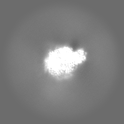
| Images | 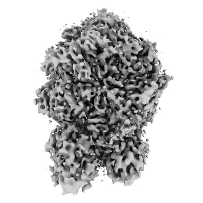 emd_41411.png emd_41411.png | 99.5 KB | ||
| Filedesc metadata | emd-41411.cif.gz | 7.3 KB | ||
| Others | emd_41411_additional_1.map.gz | 30.1 MB | ||
| Archive directory |  http://ftp.pdbj.org/pub/emdb/structures/EMD-41411ftp://ftp.pdbj.org/pub/emdb/structures/EMD-41411 http://ftp.pdbj.org/pub/emdb/structures/EMD-41411ftp://ftp.pdbj.org/pub/emdb/structures/EMD-41411 | HTTPS FTP |
-Related structure data
| Related structure data |  8tn9MC C: citing same article ( M: atomic model generated by this map |
|---|---|
| Similar structure data |


-Links
| EMDB pages | EMDB (EBI/PDBe) / EMDataResource |
|---|---|
| Related items in Molecule of the Month |


-Map
| File | Download / File: emd_41411.map.gz / Format: CCP4 / Size: 64 MB / Type: IMAGE STORED AS FLOATING POINT NUMBER (4 BYTES) | ||||||||||||||||||||||||||||||||||||
|---|---|---|---|---|---|---|---|---|---|---|---|---|---|---|---|---|---|---|---|---|---|---|---|---|---|---|---|---|---|---|---|---|---|---|---|---|---|
| Annotation | Coagulation plasma factor V - Sharpened composite map | ||||||||||||||||||||||||||||||||||||
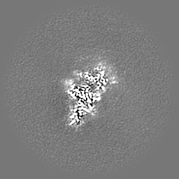
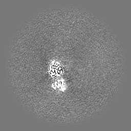
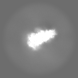
| Projections & slices | Image control
Images are generated by Spider. | ||||||||||||||||||||||||||||||||||||
| Voxel size | X=Y=Z: 1.1 Å | ||||||||||||||||||||||||||||||||||||
| Density |
| ||||||||||||||||||||||||||||||||||||
| Symmetry | Space group: 1 | ||||||||||||||||||||||||||||||||||||
| Details | EMDB XML:
|
 X (Sec.)
 Y (Row.)
 Z (Col.)


-Supplemental data
-Additional map: Coagulation plasma factor V - unsharpened composite map
| File | emd_41411_additional_1.map | ||||||||||||
|---|---|---|---|---|---|---|---|---|---|---|---|---|---|
| Annotation | Coagulation plasma factor V - unsharpened composite map | ||||||||||||
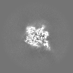
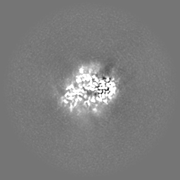
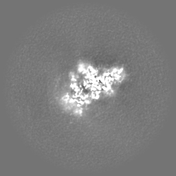
| Projections & Slices |
| ||||||||||||
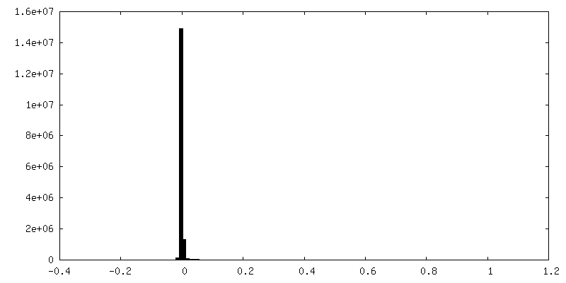
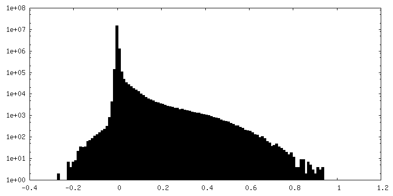
| Density Histograms |

- Sample components
-Entire : Coagulation plasma Factor V
| Entire | Name: Coagulation plasma Factor V |
|---|---|
| Components |
|
-Supramolecule #1: Coagulation plasma Factor V
| Supramolecule | Name: Coagulation plasma Factor V / type: tissue / ID: 1 / Parent: 0 / Macromolecule list: #1 |
|---|---|
| Source (natural) | Organism: Homo sapiens (human) / Tissue: Blood |
-Macromolecule #1: Coagulation factor V
| Macromolecule | Name: Coagulation factor V / type: protein_or_peptide / ID: 1 / Number of copies: 1 / Enantiomer: LEVO |
|---|---|
| Source (natural) | Organism: Homo sapiens (human) |
| Molecular weight | Theoretical: 248.973234 KDa |
| Sequence | String: AQLRQFYVAA QGISWSYRPE PTNSSLNLSV TSFKKIVYRE YEPYFKKEKP QSTISGLLGP TLYAEVGDII KVHFKNKADK PLSIHPQGI RYSKLSEGAS YLDHTFPAEK MDDAVAPGRE YTYEWSISED SGPTHDDPPC LTHIYYSHEN LIEDFNSGLI G PLLICKKG ...String: AQLRQFYVAA QGISWSYRPE PTNSSLNLSV TSFKKIVYRE YEPYFKKEKP QSTISGLLGP TLYAEVGDII KVHFKNKADK PLSIHPQGI RYSKLSEGAS YLDHTFPAEK MDDAVAPGRE YTYEWSISED SGPTHDDPPC LTHIYYSHEN LIEDFNSGLI G PLLICKKG TLTEGGTQKT FDKQIVLLFA VFDESKSWSQ SSSLMYTVNG YVNGTMPDIT VCAHDHISWH LLGMSSGPEL FS IHFNGQV LEQNHHKVSA ITLVSATSTT ANMTVGPEGK WIISSLTPKH LQAGMQAYID IKNCPKKTRN LKKITREQRR HMK RWEYFI AAEEVIWDYA PVIPANMDKK YRSQHLDNFS NQIGKHYKKV MYTQYEDESF TKHTVNPNMK EDGILGPIIR AQVR DTLKI VFKNMASRPY SIYPHGVTFS PYEDEVNSSF TSGRNNTMIR AVQPGETYTY KWNILEFDEP TENDAQCLTR PYYSD VDIM RDIASGLIGL LLICKSRSLD RRGIQRAADI EQQAVFAVFD ENKSWYLEDN INKFCENPDE VKRDDPKFYE SNIMST ING YVPESITTLG FCFDDTVQWH FCSVGTQNEI LTIHFTGHSF IYGKRHEDTL TLFPMRGESV TVTMDNVGTW MLTSMNS SP RSKKLRLKFR DVKCIPDDDE DSYEIFEPPE STVMATRKMH DRLEPEDEES DADYDYQNRL AAALGIRSFR NSSLNQEE E EFNLTALALE NGTEFVSSNT DIIVGSNYSS PSNISKFTVN NLAEPQKAPS HQQATTAGSP LRHLIGKNSV LNSSTAEHS SPYSEDPIED PLQPDVTGIR LLSLGAGEFK SQEHAKHKGP KVERDQAAKH RFSWMKLLAH KVGRHLSQDT GSPSGMRPWE DLPSQDTGS PSRMRPWKDP PSDLLLLKQS NSSKILVGRW HLASEKGSYE IIQDTDEDTA VNNWLISPQN ASRAWGESTP L ANKPGKQS GHPKFPRVRH KSLQVRQDGG KSRLKKSQFL IKTRKKKKEK HTHHAPLSPR TFHPLRSEAY NTFSERRLKH SL VLHKSNE TSLPTDLNQT LPSMDFGWIA SLPDHNQNSS NDTGQASCPP GLYQTVPPEE HYQTFPIQDP DQMHSTSDPS HRS SSPELS EMLEYDRSHK SFPTDISQMS PSSEHEVWQT VISPDLSQVT LSPELSQTNL SPDLSHTTLS PELIQRNLSP ALGQ MPISP DLSHTTLSPD LSHTTLSLDL SQTNLSPELS QTNLSPALGQ MPLSPDLSHT TLSLDFSQTN LSPELSHMTL SPELS QTNL SPALGQMPIS PDLSHTTLSL DFSQTNLSPE LSQTNLSPAL GQMPLSPDPS HTTLSLDLSQ TNLSPELSQT NLSPDL SEM PLFADLSQIP LTPDLDQMTL SPDLGETDLS PNFGQMSLSP DLSQVTLSPD ISDTTLLPDL SQISPPPDLD QIFYPSE SS QSLLLQEFNE SFPYPDLGQM PSPSSPTLND TFLSKEFNPL VIVGLSKDGT DYIEIIPKEE VQSSEDDYAE IDYVPYDD P YKTDVRTNIN SSRDPDNIAA WYLRSNNGNR RNYYIAAEEI SWDYSEFVQR ETDIEDSDDI PEDTTYKKVV FRKYLDSTF TKRDPRGEYE EHLGILGPII RAEVDDVIQV RFKNLASRPY SLHAHGLSYE KSSEGKTYED DSPEWFKEDN AVQPNSSYTY VWHATERSG PESPGSACRA WAYYSAVNPE KDIHSGLIGP LLICQKGILH KDSNMPMDMR EFVLLFMTFD EKKSWYYEKK S RSSWRLTS SEMKKSHEFH AINGMIYSLP GLKMYEQEWV RLHLLNIGGS QDIHVVHFHG QTLLENGNKQ HQLGVWPLLP GS FKTLEMK ASKPGWWLLN TEVGENQRAG MQTPFLIMDR DCRMPMGLST GIISDSQIKA SEFLGYWEPR LARLNNGGSY NAW SVEKLA AEFASKPWIQ VDMQKEVIIT GIQTQGAKHY LKSCYTTEFY VAYSSNQINW QIFKGNSTRN VMYFNGNSDA STIK ENQFD PPIVARYIRI SPTRAYNRPT LRLELQGCEV NGCSTPLGME NGKIENKQIT ASSFKKSWWG DYWEPFRARL NAQGR VNAW QAKANNNKQW LEIDLLKIKK ITAIITQGCK SLSSEMYVKS YTIHYSEQGV EWKPYRLKSS MVDKIFEGNT NTKGHV KNF FNPPIISRFI RVIPKTWNQS IALRLELFGC DIY UniProtKB: Coagulation factor V |
-Macromolecule #2: 2-acetamido-2-deoxy-beta-D-glucopyranose
| Macromolecule | Name: 2-acetamido-2-deoxy-beta-D-glucopyranose / type: ligand / ID: 2 / Number of copies: 4 / Formula: NAG |
|---|---|
| Molecular weight | Theoretical: 221.208 Da |
| Chemical component information |  ChemComp-NAG: |
-Experimental details
-Structure determination
| Method | cryo EM |
|---|---|
 Processing Processing | single particle reconstruction |
| Aggregation state | particle |
-Sample preparation
| Concentration | 0.2 mg/mL |
|---|---|
| Buffer | pH: 7.4 / Details: 20 mM HEPES, 150 mM NaCl, 5 mM CaCl2 |
| Grid | Model: Quantifoil R2/2 / Material: COPPER / Mesh: 300 / Support film - Material: CARBON / Support film - topology: HOLEY / Pretreatment - Type: PLASMA CLEANING / Pretreatment - Time: 60 sec. / Pretreatment - Atmosphere: OTHER / Pretreatment - Pressure: 0.009300000000000001 kPa |
| Vitrification | Cryogen name: ETHANE / Chamber humidity: 100 % / Chamber temperature: 277.15 K / Instrument: FEI VITROBOT MARK IV |
| Details | 0.1 and 0.2 mg/mL |
- Electron microscopy
| Microscope | FEI TITAN |
|---|---|
| Specialist optics | Energy filter - Name: GIF Bioquantum / Energy filter - Slit width: 20 eV |
| Image recording | Film or detector model: GATAN K2 SUMMIT (4k x 4k) / Detector mode: COUNTING / Digitization - Dimensions - Width: 4000 pixel / Digitization - Dimensions - Height: 4000 pixel / Number grids imaged: 2 / Number real images: 8429 / Average exposure time: 1.65 sec. / Average electron dose: 66.0 e/Å2 Details: 2 Grids imaged one at 0.1 mg/mL and the second at 0.2 mg/mL using the same acquisition parameters. |
| Electron beam | Acceleration voltage: 300 kV / Electron source:  FIELD EMISSION GUN FIELD EMISSION GUN |
| Electron optics | Illumination mode: FLOOD BEAM / Imaging mode: BRIGHT FIELD / Cs: 0.01 mm / Nominal defocus max: 2.5 µm / Nominal defocus min: 1.0 µm / Nominal magnification: 105000 |
| Sample stage | Specimen holder model: FEI TITAN KRIOS AUTOGRID HOLDER / Cooling holder cryogen: NITROGEN |